What if the Higgs boson found in 2012 is not alone but is the only sibling we have encountered so far? Scientists at CERN discovered the particle that year, and it was a major discovery because it explained how other particles acquire mass. For a long time, scientists thought this was the final piece of the puzzle.
They have a framework called the Standard Model that describes the smallest particles in everything we see. This includes electrons in atoms and light particles called photons. However, this framework does not explain everything. It does not tell us about dark matter or why the universe has so much more matter than antimatter. It is like having a map that shows only half the world.
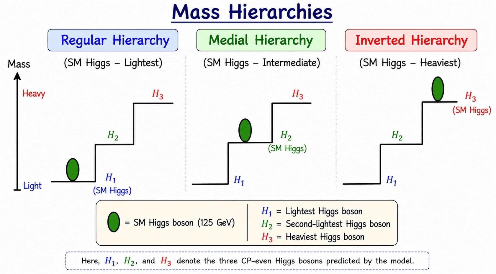
The discovery of the Higgs boson created new questions. Many physicists started wondering whether the Higgs we found is the only one of its kind. They began to ask whether there is a larger family of these particles hiding in the universe. If we find more members of this family, we might finally understand the parts of nature that the current framework misses.