We introduce Layered Self-Supervised Knowledge Distillation (LSSKD) framework for training compact deep learning models. Unlike traditional methods that rely on pre-trained teacher networks, our approach appends auxiliary classifiers to intermediate feature maps, generating diverse self-supervised knowledge and enabling one-to-one transfer across different network stages. Our method achieves an average improvement of 4.54\% over the state-of-the-art PS-KD method and a 1.14% gain over SSKD on CIFAR-100, with a 0.32% improvement on ImageNet compared to HASSKD. Experiments on Tiny ImageNet and CIFAR-100 under few-shot learning scenarios also achieve state-of-the-art results. These findings demonstrate the effectiveness of our approach in enhancing model generalization and performance without the need for large over-parameterized teacher networks. Importantly, at the inference stage, all auxiliary classifiers can be removed, yielding no extra computational cost. This makes our model suitable for deploying small language models on affordable low-computing devices. Owing to its lightweight design and adaptability, our framework is particularly suitable for multimodal sensing and cyber-physical environments that require efficient and responsive inference. LSSKD facilitates the development of intelligent agents capable of learning from limited sensory data under weak supervision.
Category: mapping
Webb maps the mysterious upper atmosphere of Uranus
For the first time, an international team of astronomers have mapped the vertical structure of Uranus’s upper atmosphere, uncovering how temperature and charged particles vary with height across the planet. Using Webb’s NIRSpec instrument, the team observed Uranus for nearly a full rotation, detecting the faint glow from molecules high above the clouds.
These unique data provide the most detailed portrait yet of where the planet’s auroras form, how they are influenced by its unusually tilted magnetic field, and how Uranus’s atmosphere has continued to cool over the past three decades. The results, published in Geophysical Research Letters, offer a new window into how ice-giant planets distribute energy in their upper layers.
Led by Paola Tiranti of Northumbria University in the United Kingdom, the study mapped out the temperature and density of ions in the atmosphere extending up to 5,000 kilometers above Uranus’s cloud tops, a region called the ionosphere where the atmosphere becomes ionized and interacts strongly with the planet’s magnetic field. The measurements show that temperatures peak between 3,000 and 4,000 kilometers, while ion densities reach their maximum around 1,000 kilometers, revealing clear longitudinal variations linked to the complex geometry of the magnetic field.

Committee co-chaired by Prof. Dava Newman issues a new roadmap for human missions to Mars
On December 9, the National Academies of Sciences, Engineering, and Medicine (NASEM) released a landmark report, A Science Strategy for the Human Exploration of Mars, laying out a comprehensive case for future crewed Mars missions. The report, authored by the Committee on a Science Strategy for the Human Exploration of Mars that was co-chaired by Prof. Dava Newman, defines the highest-priority scientific objectives for humans on the Martian surface.
At the top of the list: searching for evidence of past or present life. “We’re searching for life on Mars,” said Newman in an interview with Ars Technica. “The answer to the question ‘are we alone?’ is always going to be ‘maybe,’ unless it becomes yes.”
The report identifies 11 top science goals for initial human missions, including biosignature/habitability experiments and water and CO₂ cycle studies, geology mapping, radiation monitoring, dust-storm research, and assessments of how Martian conditions affect humans and ecosystems.

Effective connectivity between homologous cortices mediated by the corpus callosum: An axono-cortical evoked potentials study
[Functional brain mapping] Mitsuhashi et al.: “Callosal stimulation showed effective connectivity to homologous cortical regions. Sum of callosal-to-cortex propagation latencies matched interhemispheric latency.” Open access.
All content on this site: Copyright © 2026 Elsevier B.V., its licensors, and contributors. All rights are reserved, including those for text and data mining, AI training, and similar technologies. For all open access content, the relevant licensing terms apply.

Inside PC gaming’s wildly creative Tomb Raider mapping scene: ‘Being able to create my own adventures for other people to play is such an addicting concept’
“At that time, I had no professional experience in the videogame industry, and I didn’t even really have an artist portfolio, so I made one in a bit of a rush.” Nonetheless, this was enough to convince Saber, and Hatté joined the team as an environment artist, working primarily on Tomb Raider 4–6 Remastered.
“My role on the team was specifically to work on the environments and remaster the textures,” Hatté says. “It was a life changing experience. I’m incredibly grateful to have been given that opportunity.”
Reamsters aside, Tomb Raider has been dormant since 2018’s Shadow of the Tomb Raider. But late last year, two new Lara Croft adventures were revealed—a second remake of the original game, and a new adventure by Crystal Dynamics pitched as a sequel to Tomb Raider: Underworld.

Scientists Continue to Trace the Origin of the Mysterious “Amaterasu” Cosmic Ray Particle
When the Amaterasu particle entered Earth’s atmosphere, the TAP array in Utah recorded an energy level of more than 240 exa-electronvolts (EeV). Such particles are exceedingly rare and are thought to originate in some of the most extreme cosmic environments. At the time of its detection, scientists were not sure if it was a proton, a light atomic nucleus, or a heavy (iron) atomic nucleus. Research into its origin pointed toward the Local Void, a vast region of space adjacent to the Local Group that has few known galaxies or objects.
This posed a mystery for astronomers, as the region is largely devoid of sources capable of producing such energetic particles. Reconstructing the energy of cosmic-ray particles is already difficult, making the search for their sources using statistical models particularly challenging. Capel and Bourriche addressed this by combining advanced simulations with modern statistical methods (Approximate Bayesian Computation) to generate three-dimensional maps of cosmic-ray propagation and their interactions with magnetic fields in the Milky Way.
Early vertebrate biomineralization and eye structure determined by synchrotron X-ray analyses of Silurian jawless fish
Complex eyes in vertebrates may have evolved as early as 500 million years ago. Reconstructing the early evolutionary history of vertebrates requires understanding organisms that existed before bone evolved. However, these ‘soft-bodied’ fossils are inherently difficult to study, resulting in conflicting anatomical and evolutionary interpretations. For the first time, researchers used synchrotron-based imaging techniques to examine two Silurian taxa, Jamoytius and Lasanius, that may bridge the gap between the bone-less and boned vertebrates. They recover the first direct evidence of biomineralised apatitic bone in both taxa and of camera-eyes in Jamoytius. The discoveries indicate vertebrate bones and complex eyes evolved earlier than previously thought, and demonstrate the power of these techniques for problematic fossils.
Read the article in Proceedings B.
Abstract. Understanding the origin and early diversification of vertebrates has always been a challenge because of the ambiguous and conflicting interpretations of the soft-bodied, pre-biomineralization fossil record. Here, we apply synchrotron radiation techniques to Jamoytius and Lasanius, two soft-bodied Silurian vertebrates, key taxa for discerning vertebrate bone evolution owing to highly localized, but debated, biomineralization. We map soft-tissue structures and quantify details of biochemical residue impossible to resolve with traditional methods. We present the first unequivocal evidence for biomineralized apatitic scales in Jamoytius by combining synchrotron rapid scanning X-ray fluorescence and Fourier transform infrared spectroscopy (elevated Ca 37% and P 21%). This approach also recovers robust evidence for apatitic biomineralization in Lasanius. Chemical mapping of the optical anatomy of Jamoytius recovers a close correlation with Zn and Cu distribution, providing evidence for a retinal pigmented epithelium and complex eyes. In both taxa, chemical maps reveal original anatomical details not apparent in visible light, including potential evidence of other sensory anatomy in Jamoytius. Our work resolves long-standing fundamental anatomical debates, indicating stem-group origins for bone and complex eyes in vertebrates. We highlight the potential of using a powerful combination of analytical techniques to unlock otherwise inaccessible data in problematic fossils.

NOvA maps neutrino oscillations over 500 miles with 10 years of data
Neutrinos are very small, neutral subatomic particles that rarely interact with ordinary matter and are thus sometimes referred to as ghost particles. There are three known types (i.e., flavors) of neutrinos, dubbed muon, electron and tau neutrinos.
Interestingly, physicists discovered that as they travel, neutrinos can change flavor, which requires that neutrinos have a small, but not zero, mass. This phenomenon, known as neutrino oscillation, has been widely investigated in recent years, as studying it could help to infer the properties of neutrinos.
The NOvA experiment, a U.S.-based particle physics research endeavor, has been collecting data with two neutrino detectors that are far apart from each other, one located at the Fermi National Laboratory (Fermilab) in Illinois and the other at a facility in Northern Minnesota. In a recent paper, published in Physical Review Letters, the researchers involved in the NOvA experiment published some of the most precise neutrino oscillation measurements to date.


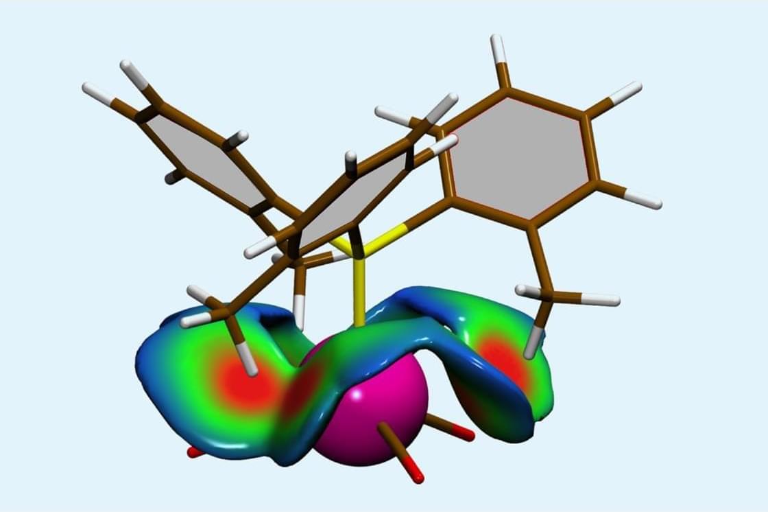
Mapping tool delivers quantitative visualisations of steric interactions
Another system the team Self on was a set of Ni–phosphine complexes, where steric interactions often limit the accessibility of the phosphine ligand’s lone pair. The researchers found that their results correlated well with the traditional method to study this – Tolman’s angles – but also showed that the interaction between the nickel centre and the ligand is smaller than Tolman analysis suggests.
These insights could prove useful for chemists. As Paton notes, ‘you could imagine comparing many different ligands in a library with this tool and using it to, for example, characterise or understand performance, maybe even ligand design… ultimately, that will allow us to think about how we might design better and more efficient systems and reactions.’
Hénon highlights that Self also works inside molecules, not just between them. ‘Previous methods were designed for two separate molecules approaching each other. But what about rotation barriers within a single molecule. That’s intramolecular steric repulsion, and Self handles it naturally. Other methods struggle badly with this. This opens new perspectives.’