The company is nearly tripling its local footprint with the move.
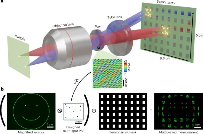
Unlike an ordinary camera, which mainly records the intensity of incoming light, a light field camera also captures information about the direction from which the light arrived. This allows it to recover depth and reconstruct a scene in three dimensions.
The technology relies on a micro-lens array (MLA) placed between the camera’s main lens and imaging sensor. Each microscopic lens acts like a tiny camera, recording the same scene from a slightly different angle. When the information from all of these lenses is combined, the system can reconstruct a light field, which describes the intensity, position, and direction of the incoming light.
Researchers have designed and built the first 3D device that can make objects invisible to heat, an advance that could transform how we protect sensitive electronics, manage heat in microchips and shield equipment from thermal detection.
The new thermal cloak can hide objects of almost any shape from infrared cameras while also protecting them from extreme temperatures. Unlike previous designs, which worked only in two dimensions or from a single direction, the cloak works from essentially any direction. Rather than simply blocking heat, thermal cloaking guides heat around an object so that, to an infrared camera, it appears as if nothing is there.
University of Illinois Urbana-Champaign civil and environmental engineering professor Shelly Zhang, postdoctoral researcher Weichen Li and graduate student Yibo Wang collaborated with professor Ole Sigmund at the Technical University of Denmark on the study, which was published in the journal Nature Communications.
When a bright fireball streaked across the Alaska sky last spring, the usual tools scientists rely on to track such events—cameras and satellites—did not provide a detailed picture. But the meteoroid left behind something else: low-frequency sound waves that traveled hundreds of miles and were captured by a dense network of earthquake and volcano-monitoring sensors on the ground.
Using those signals, a Sandia National Laboratories-led team of researchers, students and citizen scientists reconstructed the object’s path through the atmosphere, where it broke apart and where debris likely fell.
In a study published in the Journal of Geophysical Research: Planets, the team showed how low-frequency sound waves, faint ground vibrations, weather radar data and publicly shared videos can be combined to reconstruct a fireball’s path even when optical coverage is sparse or incomplete.
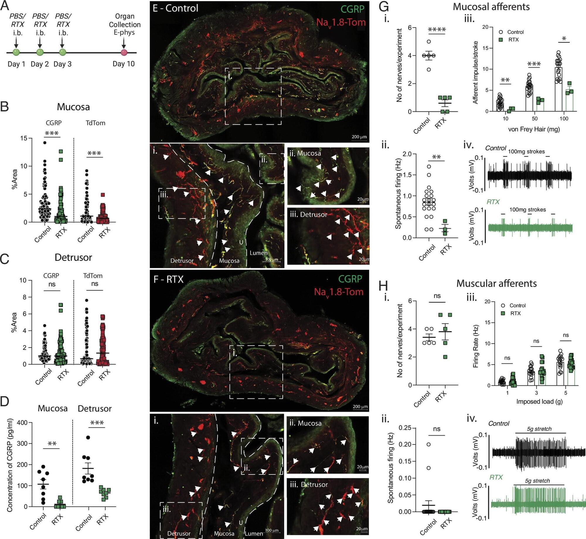
Engineers have developed a new way to monitor how tiny lab-grown human heart tissues beat—by effectively “listening” to the ripples they create. The team has created a wireless, noninvasive sensing platform that can biomechanically measure how strongly the miniature heart tissues, known as cardiac organoids, beat in real time. The research could help accelerate drug development, improve disease modeling and reduce reliance on animal testing, offering a more human-relevant way to study how the heart works.
Cardiac organoids are 3D clusters of human heart cells grown in a laboratory that are used to evaluate the safety and efficacy of new drugs prior to clinical trials, as well as study disease. While they don’t replicate the full structure of a human heart, they mimic key behaviors, especially how heart muscles contract when drugs are administered.
They are increasingly seen as a powerful alternative to animal models, which often fail to fully capture how human biology works.
To treat or manage various heart, gastrointestinal and neurological conditions, including arrhythmias, heart block, gastroparesis, epilepsy and some nerve injuries, doctors rely on a technique known as electrical stimulation. Electrical stimulation entails the delivery of small electrical pulses to target locations to prompt the activation of nerves, muscles or organs.
Many existing approaches for delivering electrical stimulation rely on electronic devices that are permanently or temporarily implanted inside the body. These devices can sometimes fail, cause adverse effects and might need to be surgically removed.
Researchers at Northwestern University, Sungkyunkwan University and other institutes recently developed a new implantable and bioresorbable system that could be used to electrically stimulate specific organs, muscles or nerves inside the body. This stimulator, presented in a paper published in Nature Electronics, could gradually disappear after a treatment is complete, so it would not need to be surgically extracted.
Europe’s first and only TES spectrometer at a synchrotron source is now in operation at BESSY II, developed within a collaboration between the HZB, the MPI-CEC (Mühlheim-an-der-Ruhr, Germany) and the NIST (Boulder, Colorado, U.S.). The photon detection efficiency of the new instrument exceeds that of wavelength-dispersive X-ray emission spectrometers by a factor of 100 to 1,000. It will be used to investigate the electronic properties of atomically thin layers, nanostructures and highly diluted atomic and molecular samples. The team is looking forward to receiving exciting research proposals from the user community.
Synchrotron radiation sources such as BESSY II provide intense, highly brilliant X-ray light that can be used to examine a wide variety of samples. However, X-ray emission spectroscopy (XES) and Resonant Inelastic X-ray Scattering (RIXS), where the photons emitted from the sample are detected, are extremely photon-hungry techniques. Therefore, XES and RIXS have so far been largely limited to high-concentration and bulk samples. The details are presented in the journal Review of Scientific Instruments.