When the engineers used gene editing to suppress the PIEZO1 gene, the cells became “deaf” to the physical tugging. Even when the magnets vigorously exercised the gel, the blood vessels barely sprouted at all. This proved that physical force directly activates this cellular gatekeeper, signaling the vessel that it’s time to grow and branch out.
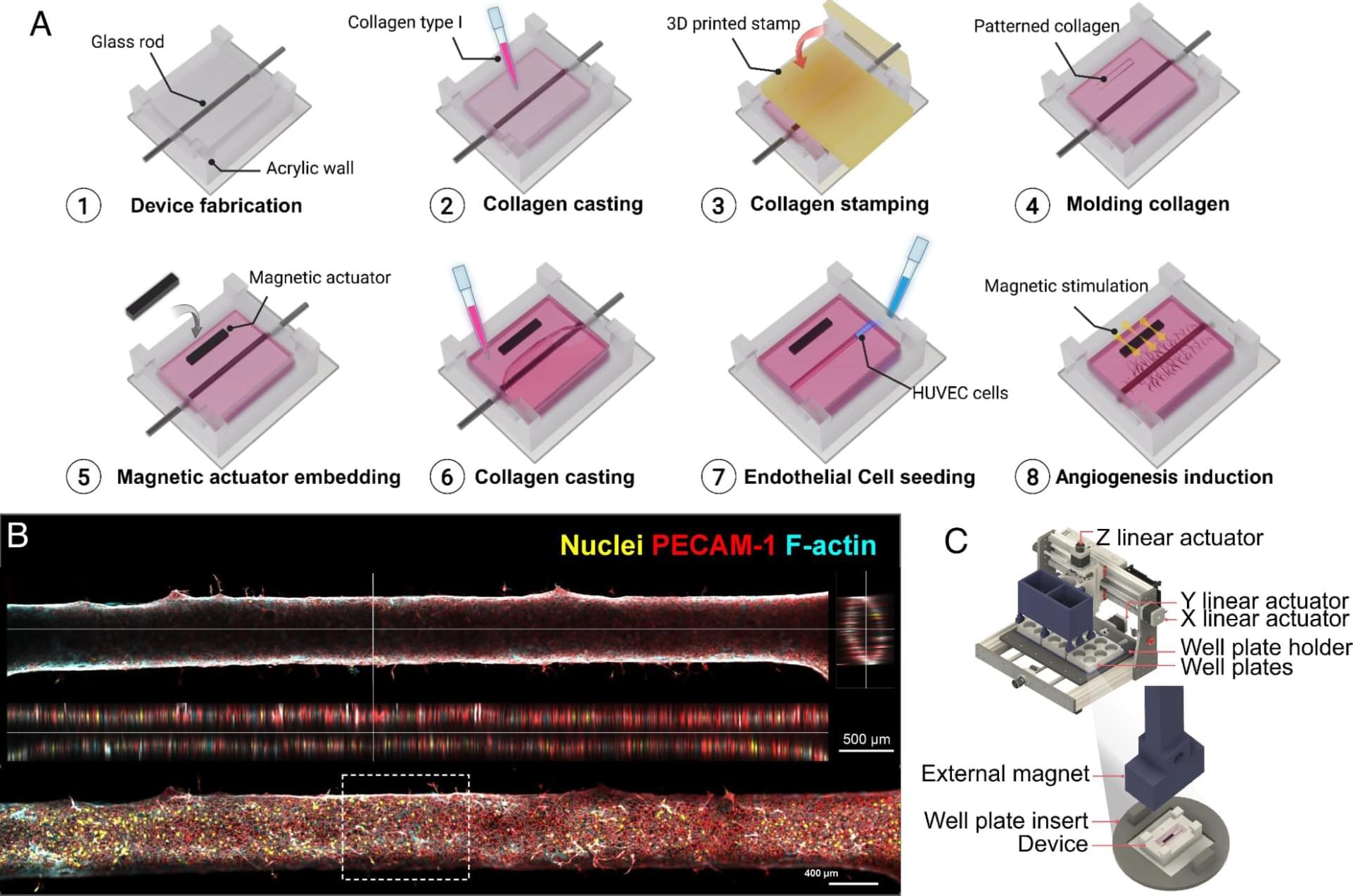
Engineering organized microvascular networks remains a critical challenge in tissue engineering and regenerative medicine. While biochemical approaches for patterning angiogenesis via growth factor delivery have shown promise, their inability to pattern sustained growth factors with spatiotemporal control limits effectiveness. Here, we demonstrate that dynamically patterned mechanical forces enable precise spatiotemporal control over angiogenic sprouting. We developed a magnetically actuated human vessel-on-a-chip platform that integrates a perfusable endothelialized microchannel within a collagen matrix and allows noninvasive and tunable mechanical stimulation across three spatial dimensions and time (4D). Using an automated 3-axis actuator, we systematically investigated how strain magnitude, frequency, and direction modulate endothelial cell behavior and vessel morphogenesis.
