Toggle light / dark theme

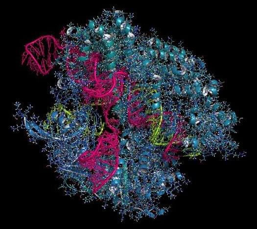
Machine learning models have seeped into the fabric of our lives, from curating playlists to explaining hard concepts in a few seconds. Beyond convenience, state-of-the-art algorithms are finding their way into modern-day medicine as a powerful potential tool. In one such advance, published in Cell Systems, Stanford researchers are using machine learning to improve the efficacy and safety of targeted cell and gene therapies by potentially using our own proteins.

Most human diseases occur due to the malfunctioning of proteins in our bodies, either systematically or locally. Naturally, introducing a new therapeutic protein to cure the one that is malfunctioning would be ideal.

Although nearly all therapeutic protein antibodies are either fully human or engineered to look human, a similar approach has yet to make its way to other therapeutic proteins, especially those that operate in cells, such as those involved in CAR-T and CRISPR-based therapies. The latter still runs the risk of triggering immune responses. To solve this problem, researchers at the Gao Lab have now turned to machine learning models.

This review explores the transformative potential of nanotechnology in the treatment and diagnosis of amyotrophic lateral sclerosis (ALS), a progressive neurodegenerative disorder characterized by motor neuron degeneration, muscle weakness, and eventual paralysis. Nanotechnology offers innovative solutions across various domains, including targeted drug delivery, neuroprotection, gene therapy and editing, biomarker detection, advanced imaging techniques, and tissue engineering. By enhancing the precision and efficacy of therapeutic interventions, nanotechnology facilitates key advancements such as crossing the blood-brain barrier, targeting specific cell types, achieving sustained therapeutic release, and enabling combination therapies tailored to the complex pathophysiology of ALS.

(Philadelphia, PA) – A single injection of a novel CRISPR gene-editing treatment safely and efficiently removes SIV – a virus related to the AIDS-causing agent HIV – from the genomes of non-human primates, scientists at the Lewis Katz School of Medicine at Temple University now report. The groundbreaking work complements previous experiments as the basis for the first-ever clinical trial of an HIV gene-editing technology in human patients, which was authorized by the Food and Drug Administration (FDA) in 2022.

The preclinical study, published online in the journal Gene Therapy, tested EBT-001, an SIV-specific CRISPR-Cas9 gene-editing therapy, in rhesus macaques. The study shows that EBT-001 effectively excises SIV from reservoirs – cells and tissues where viruses like SIV and HIV integrate into host DNA and hide for years – without any detectable off-target effects in animals. The work is a significant advance in the generation of a cure for HIV/AIDS in humans.

“Our study supports safety and demonstrates evidence of in vivo SIV editing of a CRISPR gene-editing technology aimed at the permanent inactivation of virus in a broad range of tissues in a large, preclinical animal model, using a one-time injection of the treatment,” said Kamel Khalili, PhD, Laura H. Carnell Professor and Chair of the Department of Microbiology, Immunology, and Inflammation, Director of the Center for Neurovirology and Gene Editing, Director of the Comprehensive NeuroAIDS Center at the Lewis Katz School of Medicine, and senior investigator on the new study.

A clinical study of artificial red blood cells that can be stored for transfusions in times of emergency will begin in Japan by next March, according to Nara Medical University.

The university aims to put the artificial cells into practical use by around 2030, it said in early July, in what would likely be a world first.

The development of the blood cells, designed for use in remote areas and disasters, comes as a blood shortage is expected at medical facilities due to a declining number of donors amid the country’s shrinking population.

Life on Earth possesses an exceptional ability to self-reproduce, which, even on a simple cellular level, is driven by complex biochemistry. But can self-reproduction exist in a biochemistry-free environment?

A study by researchers from Harvard University demonstrated that the answer is yes.

The researchers designed a non-biochemical system in which synthetic cell-like structures form and self-reproduce by ejecting polymeric spores.

A new, easily adopted, 3D-printed device will enable scientists to create models of human tissue with even greater control and complexity. An interdisciplinary group of researchers at the University of Washington and UW Medicine led the development of the device.

3D engineering, which recently has undergone other major advances in speed and accuracy, helps design and test therapies for a range of diseases.

One goal of tissue engineering is to create lab-made environments that recreate the natural habitats of cells.

Scientists say they’ve put together a new kind of molecular toolkit that could eventually be used to treat a variety of brain diseases, possibly including epilepsy, sleep disorders and Huntington’s disease.

The kit currently contains more than 1,000 tools of a type known as enhancer AAV vectors, with AAV standing for “adeno-associated virus.” A consortium that included researchers from Seattle’s Allen Institute for Brain Science and the University of Washington combined harmless adeno-associated viruses with snippets of engineered DNA to create a gene-therapy package that could target specific neurons in the brain while having no effect on other cells.

Researchers laid out their findings in a set of eight studies published today in the Cell Press family of journals. The work is part of a project called the Armamentarium for Precision Brain Cell Access, funded through the National Institutes of Health’s BRAIN Initiative.