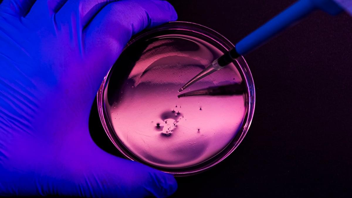
Toggle light / dark theme

Cancer Vaccines Improve Personalized Medical Care

The concept of cancer vaccines has developed over the last century with initial promise from a young doctor, William Coley. In the late 19th to early 20th century Dr. Coley developed a treatment that elicited strong immune response. This elixir was referred to as Coley’s toxin, which comprised of bacteria that generated an inflammatory response in patients. As a result, the generated response recognized and targeted the patient’s tumor. However, his treatment did not yield consistent clinical benefit. He also had his critics among physicians. At the time, the scientific community debated how safe the toxin was and whether it really worked. Colleagues at Memorial Sloan Kettering and other top institutions questioned Coley’s motive for the toxin, since there was little empirical data or scientific basis for its use. Although Coley’s toxin proved to be an inconsistent treatment, it laid the foundation for future immunotherapies as preventative and therapeutic cancer vaccines were developed.

Cancer vaccines were limited in their ability to effectively treat patients with cancer. Preventative cancer vaccines are difficult to developed because of the uncertainty to predict the onset of mutations in patients. Currently, the only U.S. Food and Drug Administration (FDA) approved preventative cancer vaccine is for the Human Papillomavirus (HPV) vaccine. While it directly protects against HPV, the vaccine indirectly prevents a multitude of cancers, including cervical, anal, and genital. Additionally, researchers have previously struggled to generate a therapeutic vaccine that elicits a strong immune response with limited adverse effects. However, a reinvigorated interest has emerged in therapeutic vaccines due to improved delivery platforms and better biomarkers to target on cancer.

Recently, an article in Cell Reports Medicine, by Dr. Nina Bhardwaj and others, examined the evolution of cancer vaccines. Specifically, the paper focused on tumor biomarker-based vaccines, which are highly personalized and designed to target genetic mutations specific to a patient’s tumor. Bhardwaj is a physician scientist, the Ward-Coleman Chair in Cancer Research, and Director of Vaccine and Cell Therapy Laboratory at the Icahn School of Medicine at Mount Sinai. Her work focuses on improving vaccine strategies to provide strong single agent affect against tumors. Bhardwaj’s group studies different cellular pathways to understand how to therapeutically target cancer.

Understanding the path from genetic changes to Parkinson’s disease opens possibilities for early diagnosis

A team led by researchers at Baylor College of Medicine and the Duncan Neurological Research Institute (Duncan NRI) at Texas Children’s Hospital has uncovered a chain of events that connects genetic alterations, disruptions in lipid metabolism and the manifestation of Parkinson’s disease in patients. The findings, published in the journal Brain, bring forward the possibility of identifying people at risk before symptoms appear and developing strategies to treat the disease rather than manage the symptoms.

“Parkinson’s disease is the second most common neurodegenerative disease after Alzheimer’s disease, affecting more than 10 million people worldwide. We know more than 100 genes that increase the risk of developing the disease but, in most cases, we do not understand how the genetic change leads to the condition,” said corresponding author Dr. Joshua Shulman, professor of neurology, neuroscience and molecular and human genetics at Baylor. He also is an investigator and co-director of the Duncan NRI.

Previous studies have shown that many Parkinson’s susceptibility genes participate in lipid metabolism and that disrupting some lipid functions may directly promote brain alterations that have been linked to the disease’s onset and progression.

Why Many Human Conceptions Don’t Last Until Birth

While estimates of total pregnancy losses vary considerably, about 15% of known pregnancies end in miscarriage, and many other conceptions do not survive past the very early stages of pregnancy. The primary cause for these losses is chromosomal abnormalities, like extra or absent chromosomes. Scientists have now analyzed data collected from over 140,000 IVF embryos to identify genetic differences that can increasethe risk of pregnancy loss. This work showed that there are certain genetic variants in some women that increase the risk of miscarriage. These findings, which were reported in Nature, may help scientists develop new methods to reduce the risk of pregnancy loss.

“This work provides the clearest evidence to date of the molecular pathways through which variable risk of chromosomal errors arises in humans,” said senior study author Rajiv McCoy, a computational biologist at Johns Hopkins University. “These insights deepen our understanding of the earliest stages of human development and open the door for future advances in reproductive genetics and fertility care.”

Rare ‘universal paralog’ genes may reveal a pre-LUCA evolutionary record

All life on Earth shares a common ancestor that lived roughly four billion years ago. This so-called “last universal common ancestor” (LUCA) represents the most ancient organism that researchers can study. Previous research on the last universal common ancestor has found that all the characteristics we see in organisms today, like having a cell membrane and a DNA genome, were already present by the time of this ancestor. So, if we want to understand how these foundational characteristics of life first emerged, then we need to be able to study evolutionary history prior to the last universal common ancestor.

In an article published in the journal Cell Genomics, scientists Aaron Goldman (Oberlin College), Greg Fournier (MIT), and Betül Kaçar (University of Wisconsin‑Madison) describe a method to do just that.

“While the last universal common ancestor is the most ancient organism we can study with evolutionary methods,” said Goldman, “some of the genes in its genome were much older.” The authors describe a type of gene family known as a “universal paralog,” which provides evidence of evolutionary events that occurred before the last universal common ancestor.

Live-cell tracking reveals dynamic interaction between protein folding helpers and newly produced proteins

Proteins are the molecular machines of cells. They are produced in protein factories called ribosomes based on their blueprint—the genetic information. Here, the basic building blocks of proteins, amino acids, are assembled into long protein chains. Like the building blocks of a machine, individual proteins must have a specific three-dimensional structure to properly fulfill their functions.

To achieve this, the newly produced protein chains in human cells are folded into their stable and functional form with the help of various protein folding helper proteins, known as chaperones, such as TRiC/PFD, or HSP70/40. The protein folding helpers isolate the amino acid chains, which have different chemical properties depending on the amino acid, from the cellular environment. This prevents the newly produced protein chains from clumping together and causing disease.

F.-Ulrich Hartl, a director at the Max Planck Institute of Biochemistry, has spent decades studying the mechanisms of protein folding. Niko Dalheimer, a scientist in Hartl’s department and one of the two lead authors of a new study published in Nature, explains: Much of what we know about protein folding has been learned from studies conducted in test tubes. However, it is virtually impossible to faithfully replicate the cellular environment in vitro.

Faster enzyme screening could cut biocatalysis bottlenecks in drug development

A team of biochemists at the University of California, Santa Cruz, has developed a faster way to identify molecules in the lab that could lead to more effective pharmaceuticals. The discovery advances the rapidly growing field of biocatalysis, which depends on generating large, genetically diverse libraries of enzymes, and then screening those variants to find ones that perform a desired chemical task best.

This strategy has attracted major investment, particularly from drugmakers, because it promises quicker routes to complex, high-value molecules. However, traditional approaches to finding new biologically beneficial molecules often require “lots of shots on goal,” where researchers test enormous numbers of candidates through slow and inefficient workflows.

The method developed by the UC Santa Cruz team aims to significantly shorten that process by introducing smarter and faster decision-making tools that help researchers identify promising enzyme variants much earlier. The researchers detail their new approach in the journal Cell Reports Physical Science.

CRISPR screen maps 250 genes essential for human muscle fiber formation

Muscles make up nearly 40% of the human body and power every move we make, from a child’s first steps to recovery after injury. For some, however, muscle development goes awry, leading to weakness, delayed motor milestones or lifelong disabilities. New research from the University of Georgia is shedding light on why.

UGA researchers have created a first-of-its-kind CRISPR screening platform for human muscle cells, identifying hundreds of genes critical to skeletal muscle formation and uncovering the potential cause of a rare genetic disorder. The findings come from two companion papers published in Nature Communications, one describing the large-scale screen and a second digging into a particular gene’s role in muscle development.

Together, the studies provide a comprehensive genetic map of how human muscle fibers are built and lend insights into the effects of genetic mutations on developmental muscle defects. By linking specific genes to the muscle-building process, this genetic roadmap gives clinicians a practical shortlist to more quickly pinpoint the likely genetic causes of a patient’s muscle-development disorder. It also provides researchers with clear targets to prioritize future drug or gene therapy approaches.

The Scientist Behind Moderna on How Engineering Revolutionizes Medicine

What does it take to turn bold ideas into life-saving medicine?

In this episode of The Big Question, we sit down with @MIT’s Dr. Robert Langer, one of the founding figures of bioengineering and among the most cited scientists in the world, to explore how engineering has reshaped modern healthcare. From early failures and rejected grants to breakthroughs that changed medicine, Langer reflects on a career built around persistence and problem-solving. His work helped lay the foundation for technologies that deliver large biological molecules, like proteins and RNA, into the body, a challenge once thought impossible. Those advances now underpin everything from targeted cancer therapies to the mRNA vaccines that transformed the COVID-19 response.

The conversation looks forward as well as back, diving into the future of medicine through engineered solutions such as artificial skin for burn victims, FDA-approved synthetic blood vessels, and organs-on-chips that mimic human biology to speed up drug testing while reducing reliance on animal models. Langer explains how nanoparticles safely carry genetic instructions into cells, how mRNA vaccines train the immune system without altering DNA, and why engineering delivery, getting the right treatment to the right place in the body, remains one of medicine’s biggest challenges. From personalized cancer vaccines to tissue engineering and rapid drug development, this episode reveals how science, persistence, and engineering come together to push the boundaries of what medicine can do next.

#Science #Medicine #Biotech #Health #LifeSciences.

Chapters:
00:00 Engineering the Future of Medicine.
01:55 Failure, Persistence, and Scientific Breakthroughs.
05:30 From Chemical Engineering to Patient Care.
08:40 Solving the Drug Delivery Problem.
11:20 Delivering Proteins, RNA, and DNA
14:10 The Origins of mRNA Technology.
17:30 How mRNA Vaccines Work.
20:40 Speed and Scale in Vaccine Development.
23:30 What mRNA Makes Possible Next.
26:10 Trust, Misinformation, and Vaccine Science.
28:50 Engineering Tissues and Organs.
31:20 Artificial Skin and Synthetic Blood Vessels.
33:40 Organs on Chips and Drug Testing.
36:10 Why Science Always Moves Forward.

The Big Question with the Museum of Science:

How DNA and life experiences leave distinct marks on the human immune system

Using single-cell epigenomic profiling of immune cells from 110 individuals, researchers show that genetic variation and environmental exposures shape the human immune system through distinct DNA methylation mechanisms. Genetic effects concentrate within gene bodies of memory cells, while environmental exposures primarily remodel regulatory regions in naive immune cells.

DNA From Ice Age Skeletons Solves a Medical Mystery That Puzzled Scientists for Decades

Scientists analyzing ancient DNA from a 12,000-year-old double burial in southern Italy uncovered genetic evidence of a rare inherited growth disorder in two closely related prehistoric individuals. A team led by researchers at the University of Vienna and Liège University Hospital Centre has tra

/* */