A study in zebrafish shows that neuronal microexons regulate brain activity by modulating cAMP signaling. Loss of these fragments leads to hyperactivity and sleep disruption, offering insights into mechanisms linked to neurodevelopmental disorders.

The first human trial of epigenetic reprogramming is underway.
Scientists are testing whether three genes can make old cells behave like young ones again while avoiding the cancer risk that has challenged the field for years.
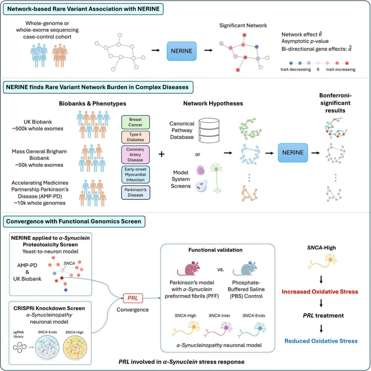
Studies of genetics conducted in yeast cells, human neurons, mice or other model systems often reveal networks of genes that could contribute to complex diseases, such as breast cancer, type 2 diabetes and Parkinson’s disease. But those findings don’t always translate to human biology. Human genetics offers a path to determining which genes among those networks are most relevant to human disease.
Researchers at Harvard Medical School have developed a new statistical framework to link networks identified in models with human genetic data. This could make it faster and easier for researchers to identify which groups of genes are most likely to contribute to a particular human disease, uncover rare disease-causing mutations and zero in on promising therapeutic targets.
The work was published in Cell Genomics.
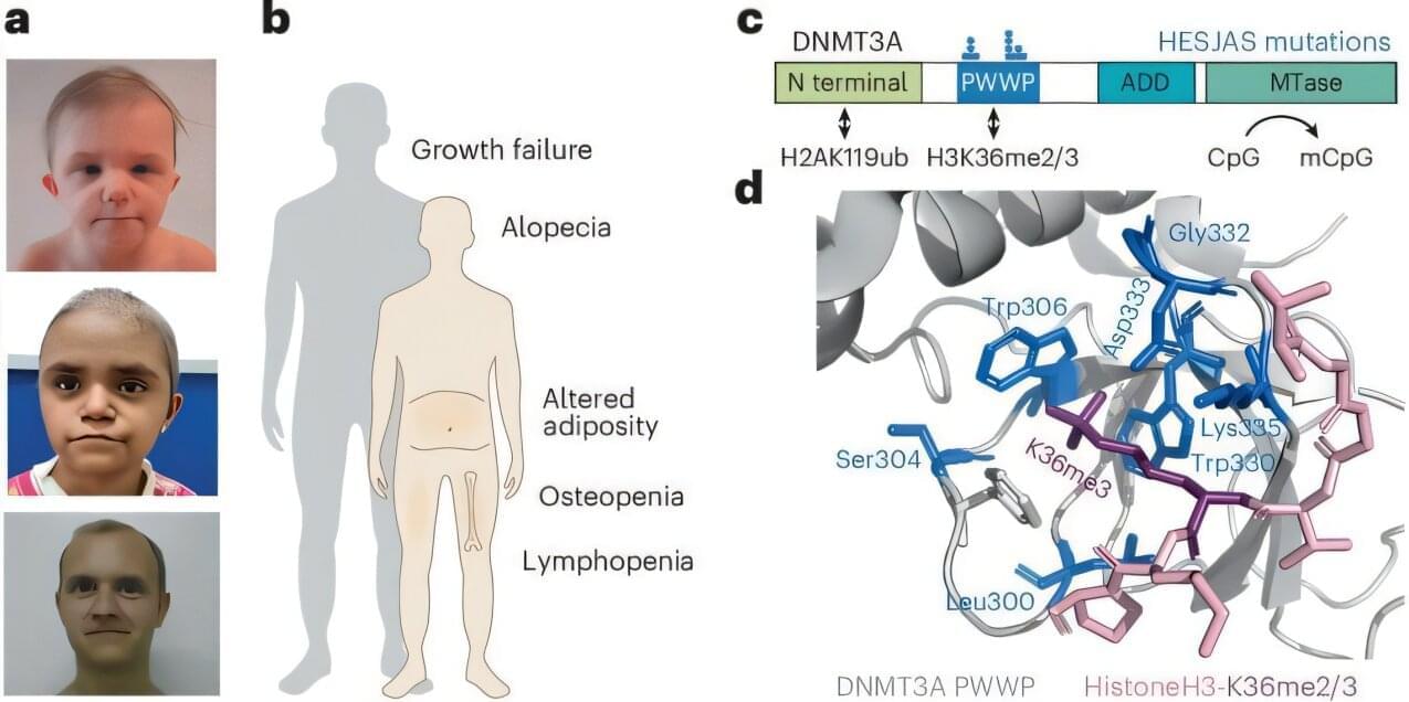
Scientists have discovered a rare genetic condition that causes people to age at a much faster rate, offering fresh insights into the aging process. The study shows for the first time how a “biological clock” present in every cell of the body could contribute to age-related diseases.
Experts say the findings could support the design of future medicines to counter diseases linked to older age, as life expectancies continue to rise across the globe.
The study is published in the journal Nature Genetics.
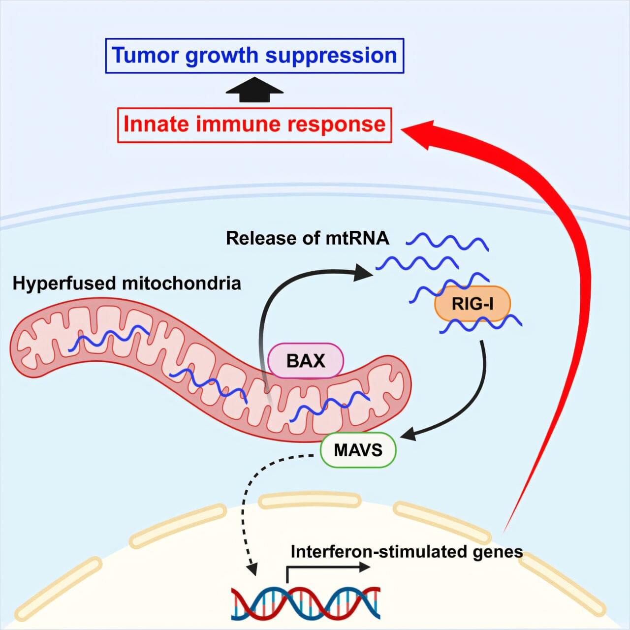
Researchers from the University of Osaka have demonstrated that mitochondrial hyperfusion, when induced by low levels of DRP1 or cellular stress, activates an immune response through the RIG-I–MAVS pathway. Dependent on the involvement of the BAX protein, the release of mitochondrial RNA into the cytosol enhanced natural killer cell cytotoxicity and reduced tumor growth in a xenograft model. The findings, published in Cell Reports, provide new possibilities for cancer research and treatment.
Mitochondria are constantly dividing and fusing within our cells, reshaping themselves to keep up with the cell’s changing needs. Sometimes, though, things go awry, and mitochondria can grow abnormally long. Are these long mitochondria harmful, or might they serve a purpose?
Mitochondria also act as signaling centers, helping the cell sense and respond to trouble. When mitochondria are hyperfused, for example in the stressed, abnormally long state described above, they release their genetic material into the cytosol, where the cell treats it as a warning sign in the same way it would treat a virus.

A compelling longitudinal study of over 350 older adults with early beta-amyloid accumulation reveals that the genetic risk for Alzheimer’s disease is not strictly deterministic, but is profoundly modulated by sleep quality through the AQP4 gene—a critical regulator of the brain’s glymphatic waste-clearance system. By cross-referencing specific AQP4 variants with multi-year MRI and PET imaging alongside cognitive assessments, researchers demonstrated that poor sleep parameters, such as shorter duration and delayed onset, significantly accelerate neurodegenerative markers like gray matter loss and ventricle expansion in carriers of specific risk alleles. Paradoxically, however, carriers of certain rare variants exhibited slower cognitive decline even in the presence of sleep disturbances. Ultimately, these findings illuminate a complex gene-environment interplay, proving that identical genetic predispositions can either expedite or buffer against brain atrophy depending on sleep architecture, thereby highlighting the critical necessity of personalized, sleep-targeted lifestyle interventions as a highly actionable strategy for Alzheimer’s prevention.
Scientists have discovered an important link between sleep, genetics, and Alzheimer’s disease. New findings suggest that getting poor sleep can accelerate brain shrinkage and memory loss in older adults carrying specific genetic variants.
Researchers from The Hospital for Sick Children (SickKids) in Toronto have found a previously unknown genetic cause of Crohn’s disease and uncovered how those changes trigger inflammation through a key immune pathway. The findings, published in Gastroenterology and involving teams from eight countries, will guide more precise treatments and improve the ability to match patients to therapies based on their unique biology.
“We’ve brought together genetics, RNA sequencing, proteomics and more to try for the first time to map the complete disease pathway, and it’s turned into a remarkable precision medicine story,” says lead author Dr. Aleixo Muise, senior scientist in the Cell & Systems Biology program, staff gastroenterologist and co-director of the Inflammatory Bowel Disease (IBD) Centre at SickKids.
“In our SickKids clinic, we want to find the right drug for each person based on their body’s unique signature. That’s why this paper is so exciting: We have pinpointed a druggable pathway.”
Most people still think Longevity Escape Velocity is a distant future. But what if some of the technologies that could make it possible are already being tested right now?
In this video, we look at three emerging longevity therapies: partial epigenetic reprogramming, senescent-cell removal, and stem-cell based repair. Some are already in human trials, while others are still early and experimental, but together they show how medicine may begin shifting from treating age-related disease to repairing parts of aging itself.
1:16 — THERAPY #1 — Partial epigenetic reprogramming.
3:34 — THERAPY #2 — SenoVax immune cleanup.
5:24 — THERAPY #3 — Lomecel — B — stem-cell therapy.
7:07 — CONCLUSION — From theory to repair.
📚 SOURCES AND STUDIES MENTIONED
ER-100 / partial epigenetic reprogramming:
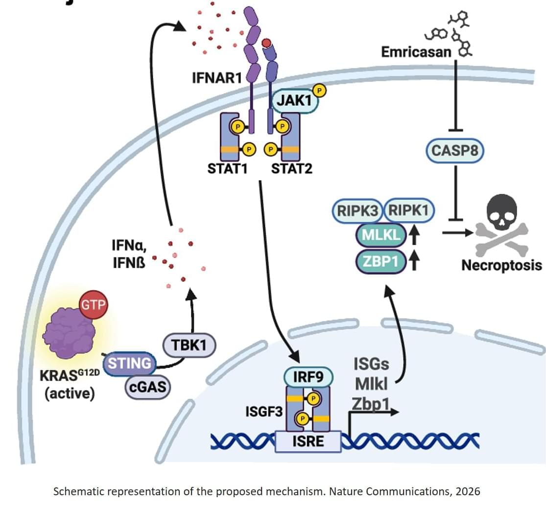
Researchers have discovered a previously unknown mechanism that makes most pancreatic cancer cells susceptible to a form of programmed cell death. The team showed that cancer cells with mutations in the KRAS gene develop a vulnerability which can be used to eliminate tumor cells in preclinical models. The findings open up new perspectives for treating pancreatic cancer. The study was published in the journal Nature Communications.
Pancreatic cancer is one of the most aggressive forms of cancer and has so far shown only limited response to available treatments. In approximately 90 percent of cases, these tumors carry mutations in the KRAS gene that drive cancer growth. Due to the ageing population and the lack of effective therapies, physicians, clinicians, and researchers expect pancreatic carcinoma to become one of the leading causes of cancer-related deaths worldwide in the coming years. With the discovery of this newly identified vulnerability, a therapeutically promising approach has now been identified for treating this disease following future clinical trials.
The researchers discovered that KRAS-mutated tumor cells continuously activate signals from the innate immune system. This primes the cancer cells for an inflammatory form of cell death known as necroptosis. In order to survive, tumor cells rely heavily on the protein caspase-8, which usually inhibits necroptosis. If caspase-8 is blocked, the tumor cells die. “KRAS-mutated tumors have a previously unknown Achilles heel,” says the senior author of the study. “By switching off the tumor cells’ defence mechanisms, we can significantly kill these tumors.”