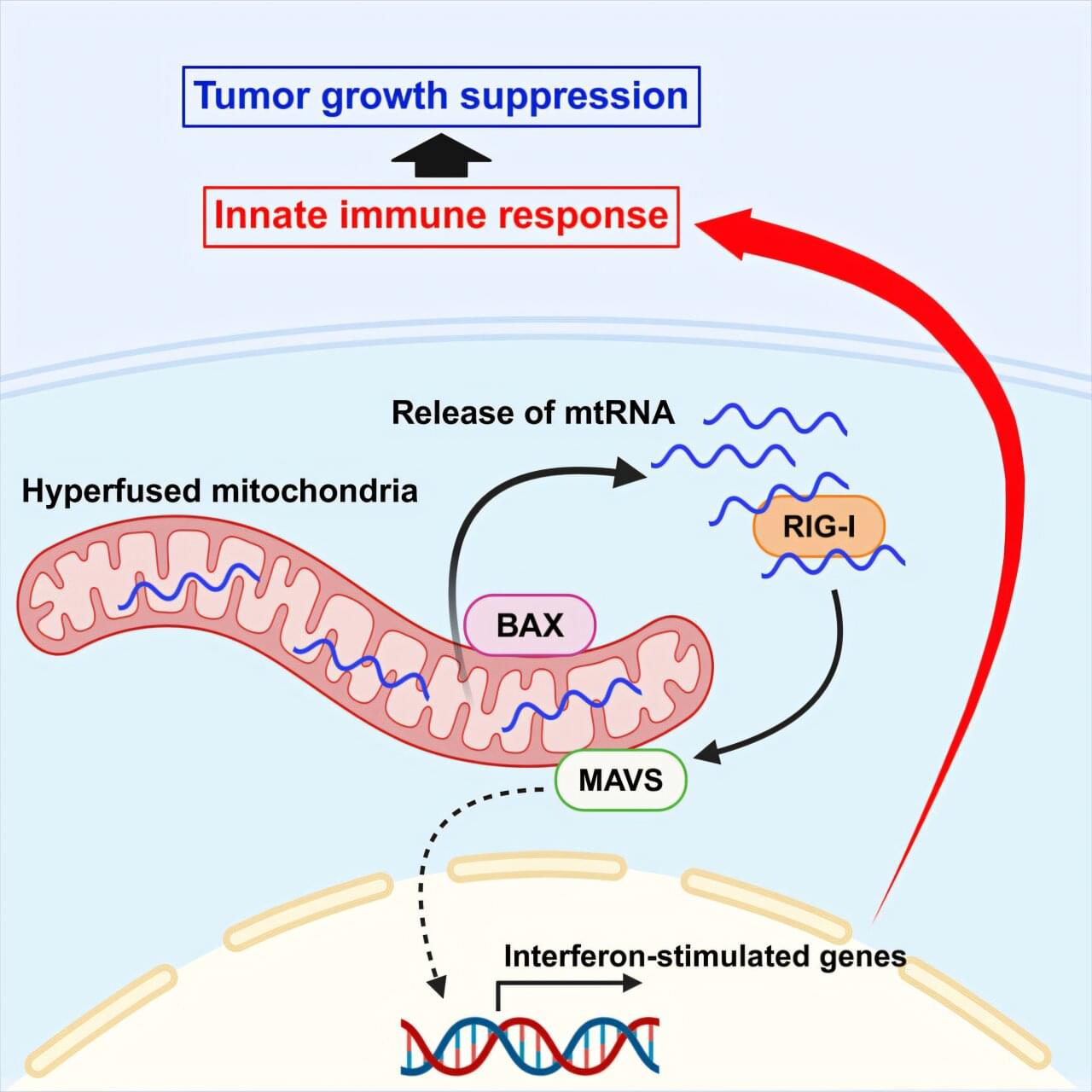
Toggle light / dark theme

A new study published in Cell Reports reveals a breakthrough discovery linking genetic variants in the gene ITSN1 to a significantly elevated risk of Parkinson’s disease, a neurodegenerative condition that affects nearly 2% of adults older than 65 years.

These findings were subsequently validated across three independent cohorts comprising more than 8,000 cases and 400,000 controls. Importantly, ITSN1 carriers trended toward earlier age of disease onset.

ITSN1 plays an important role in how neurons send messages to each other – a process called synaptic transmission – making it particularly relevant to Parkinson’s disease, a condition in which disruption of nerve signals leads to the typical symptoms of impaired gait and balance, tremors and rigidity. “We also showed in fruit flies that reducing ITSN1 levels worsens Parkinson’s-like features, including the ability to climb. We plan to extend these investigations to stem cell and mouse models,” the author said.

Interestingly, previous studies have recently implicated similar ITSN1 mutations in autism spectrum disorder (ASD). Other emerging data also have suggested an association between ASD and Parkinson’s disease, indicating that people with ASD are three times more likely to develop parkinsonism.

Ma, W., Wang, W., Zhao, L. et al. Bone Res 13, 35 (2025). https://doi.org/10.1038/s41413-025-00416-1

Download citation.

Cells function through an intricate network of proteins, each designed for specific tasks like metabolism, tissue repair, and immune defense. These proteins are built using genetic blueprints in our DNA. A process called alternative splicing enables a single gene to generate multiple mRNA transcripts — molecules carrying genetic instructions — allowing for protein diversity.

In healthy cells, this process maintains balance. Cancer cells, however, disrupt that process to fuel their unchecked growth by disabling proteins that regulate cell proliferation.

The researchers focused on a genetic element known as a poison exon. This natural “off switch” prevents the production of certain proteins by marking their RNA messages for destruction before they can be translated. Cancer cells suppress the poison exon in a key gene called TRA2β. Without this regulation, TRA2β levels rise, promoting tumor growth and making cancer cells more aggressive.

Researchers have unveiled the first real look at a mitochondrial protein strongly linked to Parkinson’s disease, revealing key details in how its malfunction might play a critical role in the disease’s progress.

Scientists have known for more than two decades that mutations in the gene for a protein called PTEN-induced putative kinase 1 (PINK1) can trigger early-onset Parkinson’s, but the mechanisms at play have remained a mystery.

A team of scientists from the Walter and Eliza Hall Institute of Medical Research (WEHI) in Australia used advanced imaging technology to not only determine the structure of PINK1, but to show how the protein attaches to cellular power houses and how they are activated.

What if everything we thought we knew about cancer was wrong?

For decades, scientists have debated what really causes cancer. Is it genetic mutations, as the Somatic Mutation Theory suggests? Is it a metabolic dysfunction, as the Metabolic Theory argues? Or is there a deeper, overlooked truth—one that could redefine cancer treatment as we know it?

In this episode, Dr. Ralph Moss and Ben Moss break down the battle between competing cancer theories, why conventional wisdom is being challenged, and what the latest research is uncovering about cancer stem cells, metabolism, and the Warburg Effect.

🔥 Are we on the verge of a breakthrough—or have we been on the wrong path all along?

📌 Subscribe for more in-depth discussions on cancer research and integrative medicine.

🔬 Resources & Further Reading:

Science, Policy And Advocacy For Impactful And Sustainable Health Ecosystems — Dr. Catharine Young, Ph.D. — fmr. Assistant Director of Cancer Moonshot Policy and International Engagement, White House Office of Science and Technology Policy (OSTP)


Dr. Catharine Young, Ph.D. recently served as Assistant Director of Cancer Moonshot Policy and International Engagement at the White House Office of Science and Technology Policy (https://www.whitehouse.gov/ostp/) where she served at OSTP to advance the Cancer Moonshot (https://www.cancer.gov/research/key-i… with a mission to decrease the number of cancer deaths by 50% over the next 25 years.

Dr. Young’s varied career has spanned a variety of sectors including academia, non-profit, biotech, and foreign government, all with a focus on advancing science.

Dr. Young previously served as Executive Director of the SHEPHERD Foundation, where she championed rare cancer research and drove critical policy changes. Her work has also included fostering interdisciplinary collaborations and advancing the use of AI, data sharing, and clinical trial reform to accelerate cancer breakthroughs.

Dr. Young’s leadership in diplomacy and innovation includes roles such as Senior Director of Science Policy at the Biden Cancer Initiative and Senior Science and Innovation Policy Advisor at the British Embassy, where she facilitated international agreements to enhance research collaborations.

The study, “Endothelial TDP-43 Depletion Disrupts Core Blood-Brain Barrier Pathways in Neurodegeneration,” was published on March 14, 2025. The lead author, Omar Moustafa Fathy, an MD/Ph. D. candidate at the Center for Vascular Biology at UConn School of Medicine, conducted the research in the laboratory of senior author Dr. Patrick A. Murphy, associate professor and newly appointed interim director of the Center for Vascular Biology. The study was carried out in collaboration with Dr. Riqiang Yan, a leading expert in Alzheimer’s disease and neurodegeneration research.

This work provides a novel and significant exploration of how vascular dysfunction contributes to neurodegenerative diseases, exemplifying the powerful collaboration between the Center for Vascular Biology and the Department of Neuroscience. While clinical evidence has long suggested that blood-brain barrier (BBB) dysfunction plays a role in neurodegeneration, the specific contribution of endothelial cells remained unclear. The BBB serves as a critical protective barrier, shielding the brain from circulating factors that could cause inflammation and dysfunction. Though multiple cell types contribute to its function, endothelial cells—the inner lining of blood vessels—are its principal component.

“It is often said in the field that ‘we are only as old as our arteries’. Across diseases we are learning the importance of the endothelium. I had no doubt the same would be true in neurodegeneration, but seeing what these cells were doing was a critical first step,” says Murphy.

Omar, Murphy, and their team tackled a key challenge: endothelial cells are rare and difficult to isolate from tissues, making it even harder to analyze the molecular pathways involved in neurodegeneration.

To overcome this, they developed an innovative approach to enrich these cells from frozen tissues stored in a large NIH-sponsored biobank. They then applied inCITE-seq, a cutting-edge method that enables direct measurement of protein-level signaling responses in single cells—marking its first-ever use in human tissues.

This breakthrough led to a striking discovery: endothelial cells from three different neurodegenerative diseases—Alzheimer’s disease (AD), amyotrophic lateral sclerosis (ALS), and frontotemporal dementia (FTD)—shared fundamental similarities that set them apart from the endothelium in healthy aging. A key finding was the depletion of TDP-43, an RNA-binding protein genetically linked to ALS-FTD and commonly disrupted in AD. Until now, research has focused primarily on neurons, but this study highlights a previously unrecognized dysfunction in endothelial cells.

“It’s easy to think of blood vessels as passive pipelines, but our findings challenge that view,” says Omar. “Across multiple neurodegenerative diseases, we see strikingly similar vascular changes, suggesting that the vasculature isn’t just collateral damage—it’s actively shaping disease progression. Recognizing these commonalities opens the door to new therapeutic possibilities that target the vasculature itself.”

One of the most enduring questions humans have is how long we’re going to live. With this comes the question of how much of our lifespan is shaped by our environment and choices, and how much is predetermined by our genes.

A study recently published in the prestigious journal Nature Medicine has attempted for the first time to quantify the relative contributions of our environment and lifestyle versus our genetics in how we age and how long we live.

The findings were striking, suggesting our environment and lifestyle play a much greater role than our genes in determining our longevity.

The landmark advance builds on a 2013 study by the team, published in Science, which described the construction of the first GRO. In that study, the researchers demonstrated new solutions for safeguarding genetically engineered organisms and for producing new classes of synthetic proteins and biomaterials with “unnatural,” or human-created, chemistries.

Ochre is a major step toward creating a non-redundant genetic code in E. coli, specifically, which is ideally suited to produce synthetic proteins containing multiple, different synthetic amino acids.

Join us on Patreon! https://www.patreon.com/MichaelLustgartenPhD

Discount Links/Affiliates:
Blood testing (where I get the majority of my labs): https://www.ultalabtests.com/partners/michaellustgarten.

At-Home Metabolomics: https://www.iollo.com?ref=michael-lustgarten.
Use Code: CONQUERAGING At Checkout.

Clearly Filtered Water Filter: https://get.aspr.app/SHoPY

Epigenetic, Telomere Testing: https://trudiagnostic.com/?irclickid=U-s3Ii2r7xyIU-LSYLyQdQ6…M0&irgwc=1
Use Code: CONQUERAGING

NAD+ Quantification: https://www.jinfiniti.com/intracellular-nad-test/