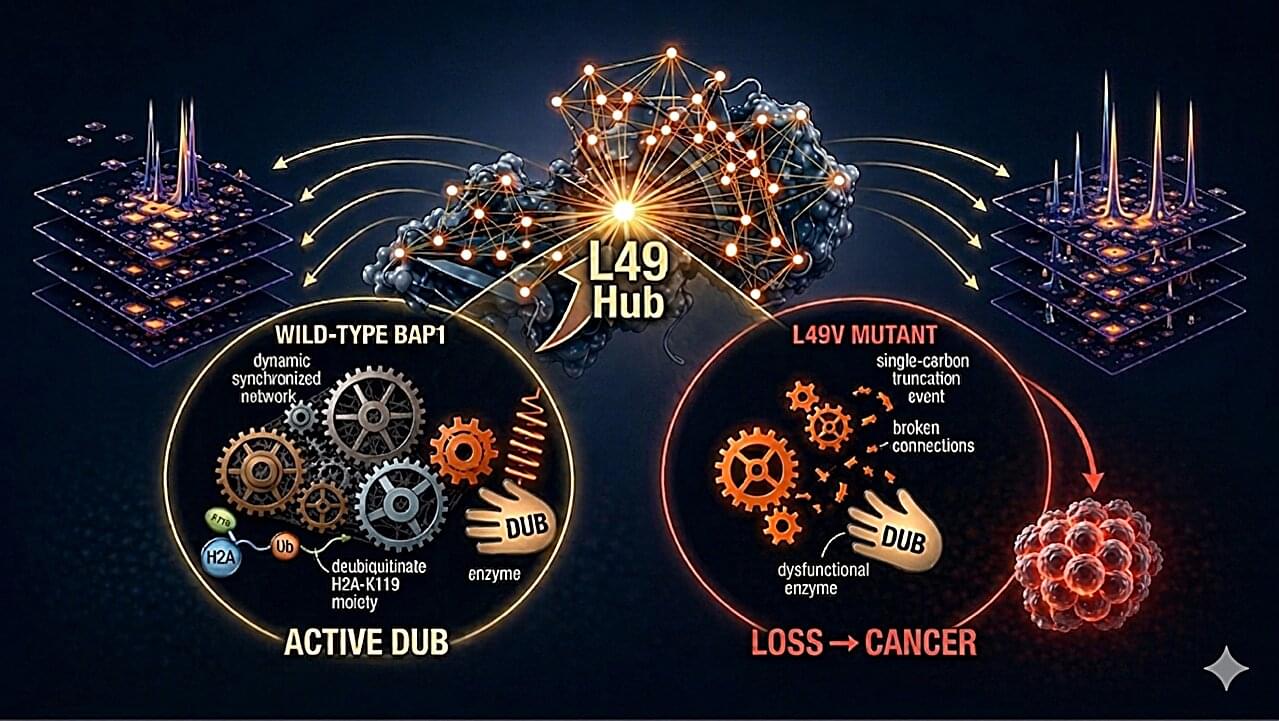
Toggle light / dark theme

New insights into the epigenetic processes via which neuroinflammation causes memory loss

Neuroinflammation, a prolonged activation of the brain’s immune system prompted by infections or other factors, has been linked to the disruption of normal mental functions. Past studies, for instance, have found that neuroinflammation plays a central role in neurodegenerative diseases, medical conditions characterized by the progressive degradation of cells in the spinal cord and brain.

When inflammation is taking place, cells release proteins that act as signals between immune cells, also known as cytokines. While some studies have linked a specific cytokine called interleukin-1 (IL-1) to changes in brain function, the mechanisms through which it could contribute to a decline in mental capabilities remain poorly understood.

Researchers at the University of Toulouse INSERM and CNRS recently carried out a study involving mice aimed at better understanding these mechanisms. Their paper, published in Nature Neuroscience, particularly focused on neuroinflammation elicited by the parasite Toxoplasma gondii (T. gondii), which is responsible for a well-known illness called toxoplasmosis.

How an autism-linked mutation reduces vasopressin and alters social behavior

A team of researchers has identified for the first time the mechanism linking a mutation in the Shank3 gene with alterations in social behavior. Using a mouse model carrying this autism-associated mutation, the study shows that vasopressin, a brain hormone essential for social relationships, is not properly released in the lateral septum.

The team is from the Cognition and Social Interactions laboratory, led by Félix Leroy at the Institute for Neurosciences, a joint center of the Spanish National Research Council (CSIC) and the Miguel Hernández University (UMH) of Elche.

The work, published in Nature Communications, demonstrates that the proper release of vasopressin in this region regulates social behaviors through two distinct receptor pathways: one controlling sociability and the other controlling social aggression, and that selective activation of these receptors can reverse deficits in social interaction without triggering unwanted aggressive responses.

Apertura Gene Therapy and Rett Syndrome Research Trust Collaborate to Pioneer Advanced Genetic Medicines for Rett Syndrome Using TfR1-Targeted AAV Capsid

NEW YORK and TRUMBULL, Conn., April 30, 2025 /PRNewswire/ — Apertura Gene Therapy, a biotechnology company focused on innovative gene therapy solutions, and the Rett Syndrome Research Trust (RSRT), an organization working to cure Rett Syndrome, today announced a collaboration to license Apertura’s human transferrin receptor 1 capsid (TfR1 CapX). This partnership aims to advance innovative genetic medicine approaches for the treatment of Rett Syndrome, a rare genetic neurological disorder caused by random mutations in the MECP2 gene on the X chromosome that primarily affect females, causing developmental regression and severe motor and language impairments.

Apertura’s TfR1 CapX is an intravenously delivered adeno-associated virus (AAV) capsid engineered to bind the transferrin receptor 1(TfR1), enabling efficient delivery of genetic medicines across the blood-brain barrier (BBB). TfR1 is a well-characterized BBB-crossing receptor, broadly and consistently expressed throughout life—even in the context of neurological disease—making it an attractive target for CNS delivery in disorders like Rett syndrome. Developed by Apertura’s academic founder, Dr. Ben Deverman, Director of Vector Engineering at the Broad Institute, TfR1 CapX has shown strong CNS selectivity in preclinical studies, achieving over 50% neuronal and 90% astrocyte transduction across multiple brain regions. Because Rett syndrome affects the brain diffusely, broader cellular transduction may correlate with greater symptomatic improvement.

Memory consolidation requires reactivation of only three neurons during sleep, research reveals

Researchers at Tsukuba University in Japan report that memories acquired while awake are stored in a more permanent form (called memory consolidation) during the REM stage of sleep, and that this process requires the reactivation of only a few specialized neurons involved in memory formation. They found that three of these neurons are crucial for memory consolidation during REM sleep.

The researchers focused on adult-born (ABNs) in the hippocampal region of the temporal lobe, which are rare neurons known to be essential for maintaining proper memory function as the loss of these cells is observed in Alzheimer’s disease. However, it has remained unclear why the loss of this small neuronal population has such devastating effects on memory.

In the Nature Communications study, specially genetically modified , in which the activity of ABNs could be monitored, were exposed to a fear experience, and the researchers examined if the activities of these ABNs during initial memory formation were reproduced during REM sleep, when dreaming is believed to occur.

New AI model predicts which genetic mutations truly drive disease

Scientists at Mount Sinai have created an artificial intelligence system that can predict how likely rare genetic mutations are to actually cause disease. By combining machine learning with millions of electronic health records and routine lab tests like cholesterol or kidney function, the system produces “ML penetrance” scores that place genetic risk on a spectrum rather than a simple yes/no. Some variants once thought dangerous showed little real-world impact, while others previously labeled uncertain revealed strong disease links.

Less is more: Gene loss drives adaptive evolution of a pandemic bacterium

A study published in Nature Ecology & Evolution reveals a surprising evolutionary insight: sometimes, losing genes rather than gaining them can help bacterial pathogens survive and thrive.

The study was conducted by a group of scientists and coordinated by Jaime Martínez Urtaza, from the Department of Genetics and Microbiology of the Universitat Autònoma de Barcelona (UAB); Yang Chao and Falush Daniel, from the Shanghai Institute of Immunity and Infection, Chinese Academy of Science; and Wang Hui, from the Shanghai Jiao Tong University.

When we think of evolution, we often imagine organisms changing or gaining to adapt, such as growing wings, developing resistance, or evolving new behaviors. Across the tree of life, both spontaneous mutations and gene acquisition are classic tools of adaptation. However, in this study, researchers went down a lesser known and scarcely explored evolutionary path, the one of gene loss.

AI and lab tests to predict genetic disease risk

When genetic testing reveals a rare DNA mutation, doctors and patients are frequently left in the dark about what it actually means. Now, researchers have developed a powerful new way to determine whether a patient with a mutation is likely to actually develop disease, a concept known in genetics as penetrance.

The team set out to solve this problem using artificial intelligence (AI) and routine lab tests like cholesterol, blood counts, and kidney function. Details of the findings were reported in the journal Science. Their new method combines machine learning with electronic health records to offer a more accurate, data-driven view of genetic risk.

Traditional genetic studies often rely on a simple yes/no diagnosis to classify patients. But many diseases, like high blood pressure, diabetes, or cancer, don’t fit neatly into binary categories. The researchers trained AI models to quantify disease on a spectrum, offering more nuanced insight into how disease risk plays out in real life.

Using more than 1 million electronic health records, the researchers built AI models for 10 common diseases. They then applied these models to people known to have rare genetic variants, generating a score between 0 and 1 that reflects the likelihood of developing the disease.

A higher score, closer to 1, suggests a variant may be more likely to contribute to disease, while a lower score indicates minimal or no risk. The team calculated “ML penetrance” scores for more than 1,600 genetic variants.

Some of the results were surprising, say the investigators. Variants previously labeled as “uncertain” showed clear disease signals, while others thought to cause disease had little effect in real-world data.

Direct plasma membrane-to-ER lipid transfer outpaces vesicular trafficking, study reveals

Max Planck Institute of Molecular Cell Biology and Genetics led a study showing that directional, non-vesicular lipid transport drives fast, species-selective lipid sorting, outpacing slower, less specific vesicular trafficking, and yielding a quantitative map of retrograde lipid transport in cells.

Thousands of lipid species occupy distinct organelle membranes, with task differences that determine cellular function. Gaps in live-cell imaging capabilities have limited clarity on how individual lipids move between organelles to maintain those tasks.

Biosynthesis of lipids begins in the (ER), followed by distribution toward the and subsequent recycling back into the ER or catabolism in lysosomes, peroxisomes, and mitochondria.

CRISPR’s efficiency triples in lab tests with DNA-wrapped nanoparticles

With the power to rewrite the genetic code underlying countless diseases, CRISPR holds immense promise to revolutionize medicine. But until scientists can deliver its gene-editing machinery safely and efficiently into relevant cells and tissues, that promise will remain out of reach.

Now, Northwestern University chemists have unveiled a new type of nanostructure that dramatically improves CRISPR delivery and potentially extends its scope of utility.

Called lipid nanoparticle spherical nucleic acids (LNP-SNAs), these tiny structures carry the full set of CRISPR editing tools—Cas9 enzymes, guide RNA and a DNA repair template—wrapped in a dense, protective shell of DNA. Not only does this DNA coating shield its cargo, but it also dictates which organs and tissues the LNP-SNAs travel to and makes it easier for them to enter cells.

/* */