Toggle light / dark theme

New sensor sniffs out pneumonia on a patient’s breath

Diagnosing some diseases could be as easy as breathing into a tube. MIT engineers have developed a test to detect disease-related compounds in a patient’s breath. The new test could provide a faster way to diagnose pneumonia and other lung conditions. Rather than sit for a chest X-ray or wait hours for a lab result, a patient may one day take a breath test and get a diagnosis within minutes.

The new breath test is a portable, chip-scale sensor that traps and detects synthetic compounds, or “biomarkers,” of disease, which are initially attached to inhalable nanoparticles. The biomarkers serve as tiny tags that can only be unlocked and detached from the nanoparticle by a very particular key, such as a disease-related enzyme.

The idea is that a person would first breathe in the nanoparticles, similar to inhaling asthma medicine. If the person is healthy, the nanoparticles would eventually circulate out of the body intact. If a disease such as pneumonia is present, however, enzymes produced as a result of the infection would snip off the nanoparticles’ biomarkers. These untethered biomarkers would be exhaled and measured, confirming the presence of the disease.

Dynamic interactions between brain tumors and immune cells

Glioblastoma, the most common and most aggressive brain tumor type in adults, remains difficult to treat because it can infiltrate surrounding brain tissue and spread far beyond the main tumor. Researchers have captured this infiltration process in the living brain with advanced microscopy. Their study is based on observations in mice affected by a brain cancer very similar to human glioblastoma.

The results, published in the scientific journal Immunity, reveal complex and situation-dependent interactions between glioblastoma cells and the brain’s resident immune cells, also known as “microglia”. These cells patrol the brain in search of threats. The current findings suggest that microglia are not passive bystanders, but actively influence both the containment and the spread of the tumor.

The scientists observed these processes by means of so-called three-photon microscopy that employs infrared light. Focus was on the “far infiltration zone”, which designates a tissue region located several millimeters away from the primary tumor.

Among other things, the team discovered that the behavior of microglia changed as a tumor spread. Specifically, microglia showed increased motility and surveillance activity when only a few glioblastoma cells were present. However, as tumor infiltration intensified, this immune response declined.

Besides, the scientists investigated the effects of disabling a certain receptor that microglia use to sense their environment. The authors show that CX3CR1 deficiency enhanced microglial reactivity while limiting GB cell migration.

Furthermore, they looked into pharmacological depletion, i.e., drastically reducing the number of immune cells. Microglia depletion with the CSF1R inhibitor PLX5622 reduced GB cell migration and constrained tumor microtube ™ plasticity. ScienceMission sciencenewshighlights.

Could a natural hormone reverse obesity? New research reveals the answer

In a groundbreaking study, scientists have unlocked a major piece of the obesity puzzle, discovering that a naturally occurring hormone can reverse weight gain by targeting the same control center in the brain as popular weight-loss drugs like Ozempic and Wegovy.

The study, led by researchers at the University of Oklahoma and published in Cell Reports, highlighted the hormone FGF21 as a powerful tool in regulating metabolism and appetite.

For years, scientists assumed that weight-regulating signals primarily targeted the hypothalamus. However, Dr. Matthew Potthoff and his team were surprised to find that FGF21 actually bypasses that area, sending signals instead to the hindbrain-the lower back portion of the brain.

“Micro-managing” immune activation and protein turnover: microglial lysosomes in the context of health and disease

Microglial lysosomes immune activation and protein turnover.

In addition to its role in protein and organelle homeostasis, lysosomes are also involved in nutrient sensing, cell metabolism, immune response, and programmed cell death.

Lysosomes are heterogeneous subpopulations and their dysfunction has been associated with the pathogenesis of several neurodegenerative diseases.

Although lysosomal biogenesis, transport, and heterogeneity are well studied in neurons, the researchers in this review discuss microglial lysosome biology its regulation, composition, and function, and how these properties are linked to immune activation, aging, and certain disease pathologies. sciencenewshighlights Science Mission https://sciencemission.com/microglial-lysosomes


Npj Dementia — “Micro-managing” immune activation and protein turnover: microglial lysosomes in the context of health and disease. npj Dement. 2, 35 (2026). https://doi.org/10.1038/s44400-026-00086-8

Protein interactions in childhood brain cancer

Neuroblastoma is an unusual tumor disease of the nervous system that almost exclusively affects children, mainly younger than two years old. About half of the children have high-risk tumors with a lower chance of being cured. N-MYC is linked to poorer prognosis in neuroblastoma.

Most proteins have a definite three-dimensional structure that usually contributes to their function and how they interact with other proteins. MYC is different and does not really have a fixed three-dimensional structure. The protein is flexible and constantly changes shape, which poses a challenge to researchers seeking to understand how MYC proteins work.

Also, MYC proteins are involved in the processes necessary for healthy cells to grow and divide. To prevent all cells in the body being harmed, it is important that a drug inhibits only the MYC function that is the problem in cancer cells, and nothing else. In other words, it takes a molecule that specifically affects a certain interaction between N-MYC and another protein.

In the current study, the researchers focused on the protein Aurora A, which also has a role in neuroblastoma and many other tumor forms. Preventing these proteins from interacting with each other has been suggested as a way to treat childhood tumors.

“To stop an interaction, you need to know where it’s happening. Despite the fact that N-MYC constantly changes shape, we now know where the two proteins anchor to each other. This provided clues as to what the medication should look like. We’ve also found a small molecule that manages to break apart the proteins, which lays a good foundation for future clinical trials,” says the first author.

The authors show that N-Myc binding to the Aurora A N-lobe can be inhibited by the small-molecule AurkinA, providing opportunity for therapeutical strategies to disrupt this interaction. ScienceMission sciencenewshighlights.


Russia Develops ‘Anti-Aging Vaccine’ Targeting Cellular Aging

Russia is developing what officials have described as a “vaccine against aging,” a gene therapy drug aimed at slowing cellular aging by blocking a receptor linked to age-related changes in the body, the deputy science minister said Thursday.

Denis Sekirinsky, Russia’s deputy science and higher education minister, said the experimental treatment would target the RAGE receptor, which he said triggers cellular aging when activated.

“The RAGE gene is a receptor whose activation launches the aging of the cell. Blocking this gene, on the contrary, can prolong its youth,” Sekirinsky said at a healthy longevity conference in the Volga city of Saransk, according to the state-run TASS news agency.

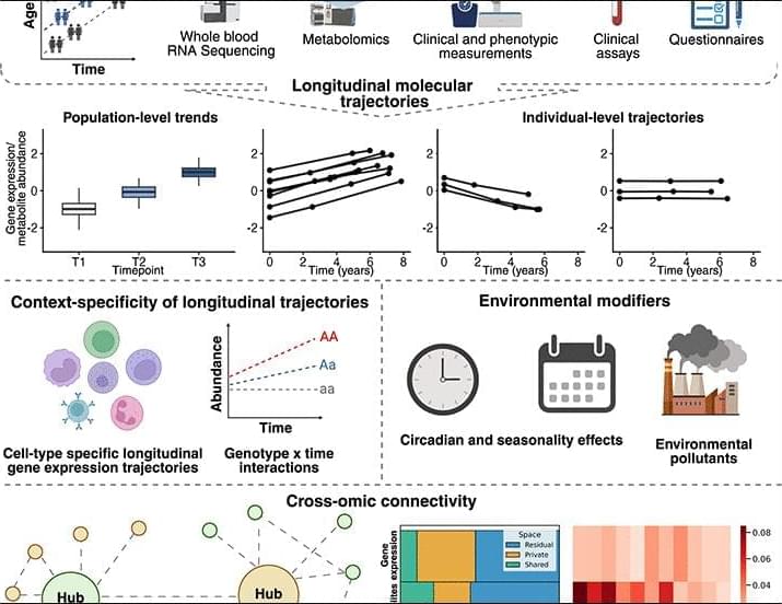
Women’s immune systems show bigger age-related changes than men’s, study reveals

Statistics show clear differences in the population’s immune system according to sex: men are more susceptible to infections and cancers, while women have stronger immune responses, which translate, for example, into better responses to vaccines. Even so, with a more reactive immune system, the probability of the body attacking itself also increases, causing 80% of autoimmune disease development to occur in women.

In this context, understanding the aging of the immune system is key since, with age, the composition of immune cells changes and their protective functions deteriorate, causing a greater susceptibility to diseases. However, understanding how sex influences this profound transformation was not possible until now.

A new study by the Barcelona Supercomputing Center—Centro Nacional de Supercomputación (BSC-CNS) published today in Nature Aging demonstrated, for the first time, that immunological aging follows different dynamics between men and women, identifying the cells and genes responsible for the process, and providing a molecular explanation for the differences that previously were only observed globally in the population.

The antimetastatic effects of calorie restriction are negated by voluntary exercise in an aggressive breast cancer mouse model

The role of lifestyle interventions in treatment success has become essential for nearly every disease. Healthy dietary habits, regular exercise, and stress management are key pillars that can improve quality of life during treatment, as well as delay disease onset and progression. In this study, we focus on the combination of mild calorie restriction (CR) and voluntary exercise as coadjuvants to chemotherapy in the treatment of triple-negative breast cancer using the 4T1 mouse model. In this model, voluntary exercise did not add benefits beyond chemotherapy plus CR in terms of primary tumor size, body composition, or physical performance, while dampening the antimetastatic effect of CR in the lungs of sedentary mice. These findings highlight the challenges of translating results from one preclinical model to another, and ultimately to humans.

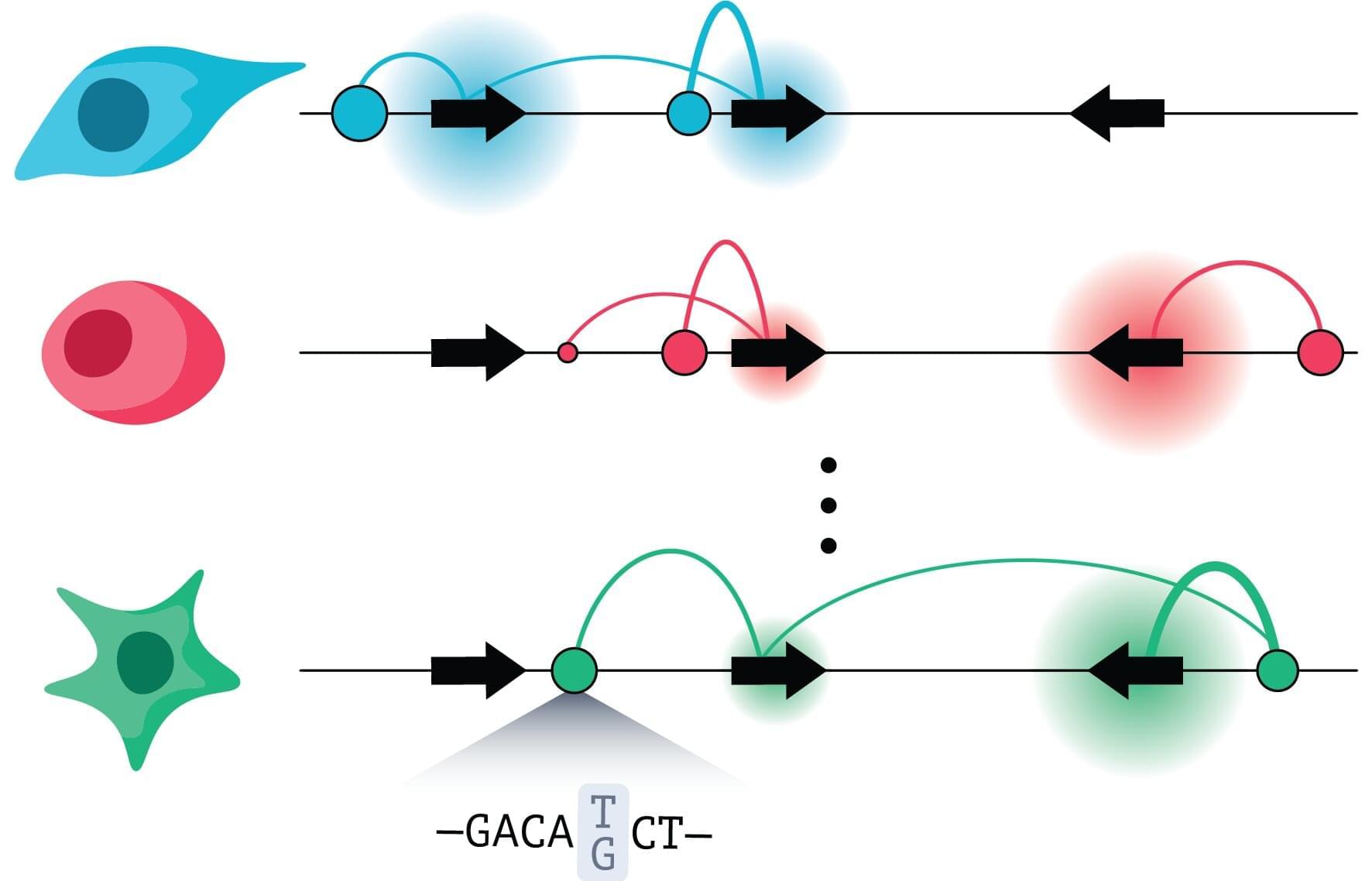
Cell-by-cell analysis uncovers 345 risk genes across six neuropsychiatric disorders

The emergence of neuropsychiatric disorders, conditions that affect various brain functions and behaviors, is known to be driven by an intricate combination of factors. These can include both a genetic predisposition and exposure to traumatic events or other external circumstances.

Over the past decades, many neuroscience studies have tried to shed light on the origins of different mental health disorders. However, the biological, cellular and molecular mechanisms underpinning these disorders have not yet been clearly elucidated.

Researchers at Peking University Sixth Hospital and Peking University Institute of Mental Health recently analyzed genetic data collected from patients diagnosed with six different neuropsychiatric disorders, to better delineate the genes and cell types that contribute to their emergence. Their paper, published in Molecular Psychiatry, identifies 345 genes expressed in different types of cells that were linked to an increased risk of developing these disorders.

/* */