Toggle light / dark theme

Bonn researchers successfully rejuvenate human cells in a test tube

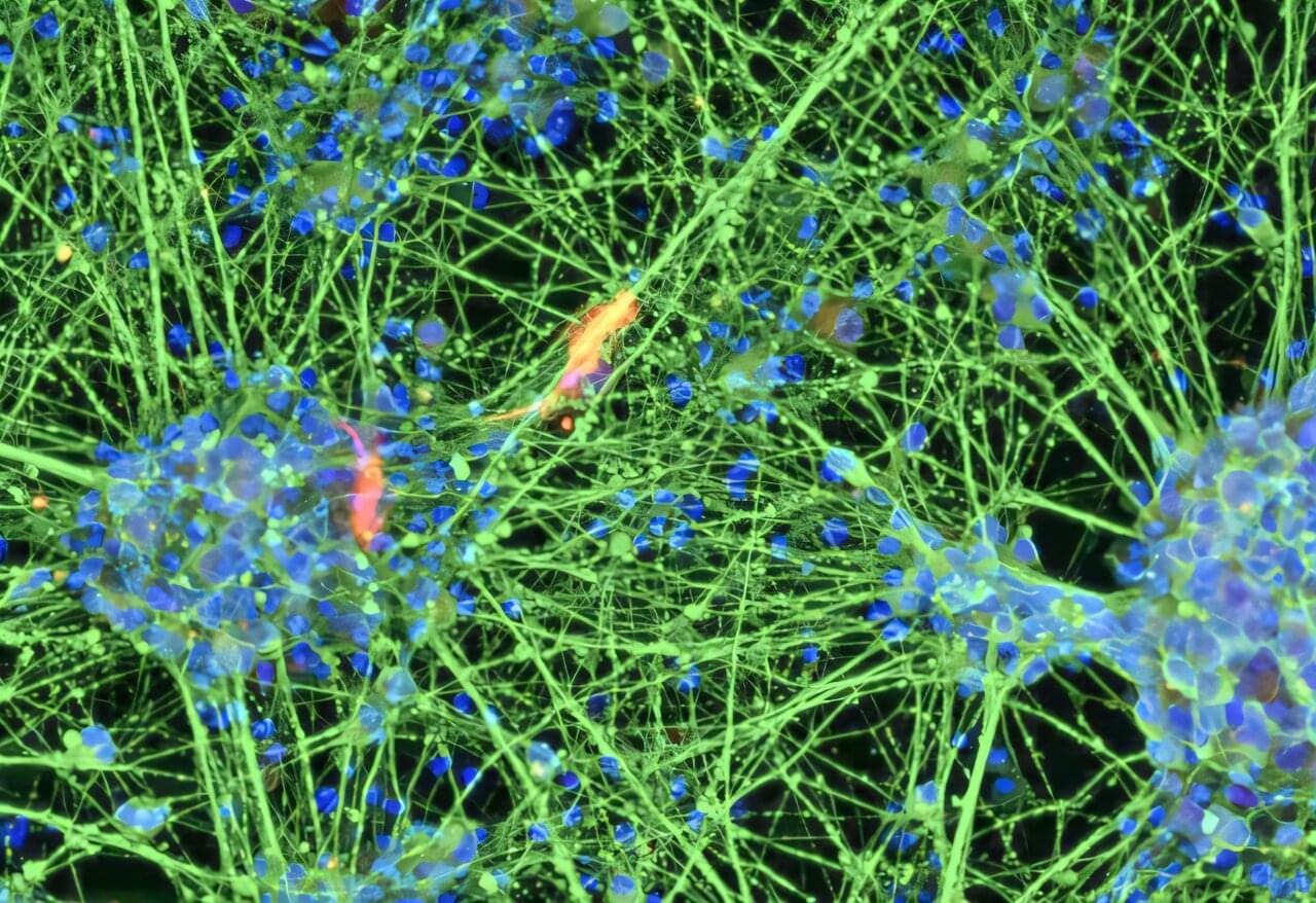
Researchers from the University Hospital of Bonn and the University of Bonn have succeeded in dramatically rejuvenating human cells in a test tube. To do so, they directly reprogrammed red blood cell precursors into stem cells from which neurons can be derived. During this process, the molecular clocks that indicate a cell’s age were reset. For example, blood cells from an 80-year-old were transformed into stem cells with a molecular age of less than 20 years. Since this rejuvenation process occurred very slowly, it is well-suited for a more detailed investigation of the underlying mechanisms. The results have been published in the journal Aging Cell.

The human body contains hundreds of different cell types. They all originate from the fertilized egg and therefore share the same genetic makeup. However, in the course of their development, they were committed to a specific fate: a skin cell cannot naturally become a liver cell, nor can a blood cell become a nerve cell.

Today, however, it is possible to reverse this determination in the laboratory. For example, nerve tissue can be grown from a skin cell, which may one day be used to treat neurodegenerative diseases. To achieve this reprogramming, researchers use a cocktail of various transcription factors. These factors cause the cell to read different genetic instructions and thus embark on a different developmental path.
The Steel Was Never Real

7 review: how falling walls between science, bodies, and AI are changing everything.

https://youtu.be/tNXNZLZI7Us](https://youtu.be/tNXNZLZI7Us)

*Oct 10, 2026 • S7 E75 • 50:52*

What if the walls between the sciences were never real? 🗄️🔥

After 75 deep dives, Heliox found a hidden pattern: the best answers live in the overlap between disciplines.

In “The Steel Was Never Real,” you’ll hear:

• How a Bayesian model revealed that two identical heart attacks can have completely different biological causes.

Ultrathin mesh harvests energy from human heart cells to generate electricity

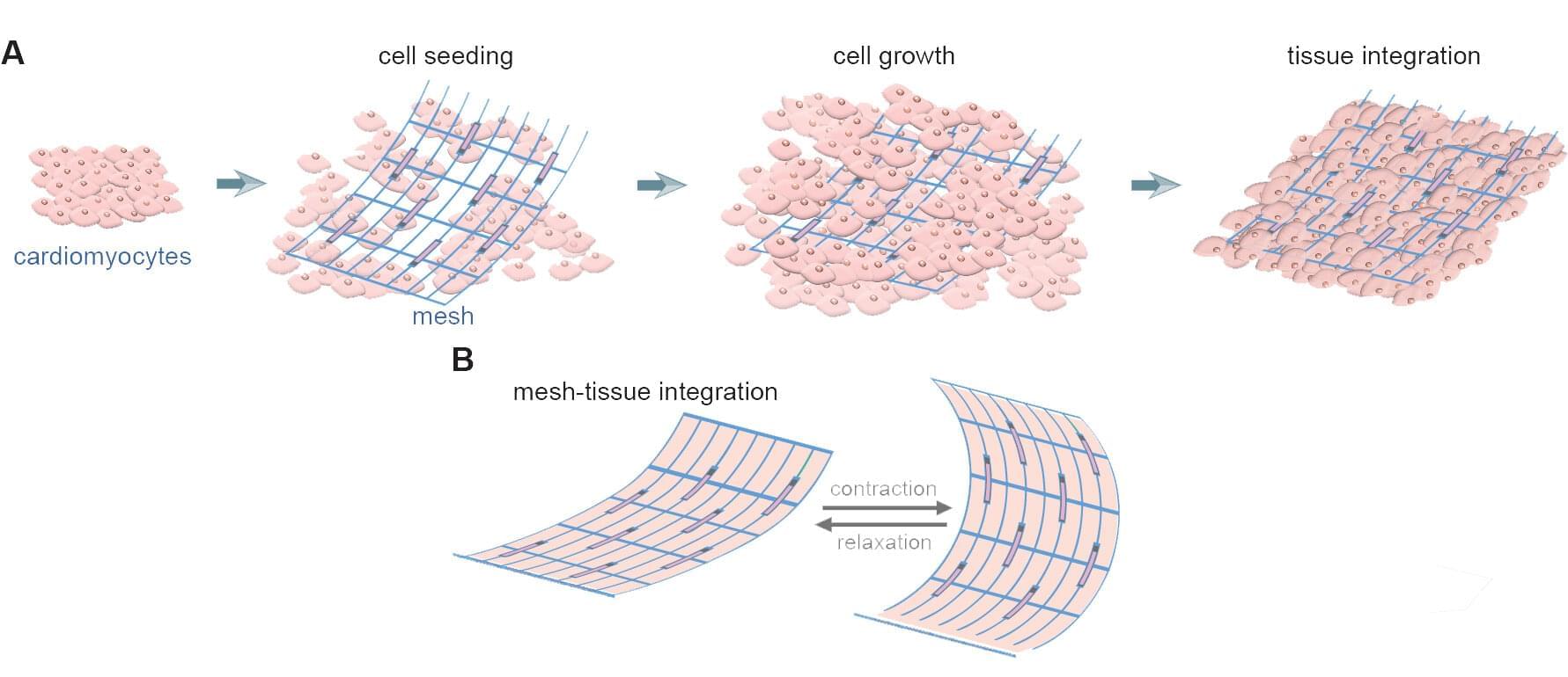
A team of engineers led by the University of Massachusetts Amherst has designed an ultrathin, flexible mesh that seamlessly integrates electronics with human cells to provide a continuous, reliable, powerful electrical supply. The study, published in Science Advances, provides a potential solution to one of the most difficult problems in wearable and implantable electronics: how to get rid of batteries.

“Humans have long dreamed of a future where certain electronics can augment our abilities,” says Jun Yao, associate professor in UMass Amherst’s Riccio College of Engineering and the paper’s senior author. One can think of famous examples from science fiction’s cyborgs, but there are also plenty of everyday examples that have nothing to do with Star Trek: pacemakers and implantable defibrillators, for example, or deep brain stimulators, cochlear implants and various health monitors. Every one of these needs a power source.

Unfortunately, batteries—the dominant power source—are bulky and eventually run out of juice, and making them smaller and more flexible reduces the charge they can store.

Scientists discover a hidden switch that shuts down inflammation

Inflammation is one of the immune system’s most important defense responses. It helps the body fight infection and repair damaged tissue. But if that response remains active for too long, it can begin to harm healthy tissue and contribute to conditions including arthritis, heart disease, and diabetes.

Scientists have long known a great deal about how inflammation begins. What has been less clear is how the body decides that the danger has passed and shifts from fighting a threat to repairing the damage.

Scientists identify the neuroblastoma cells that signal poor outcomes

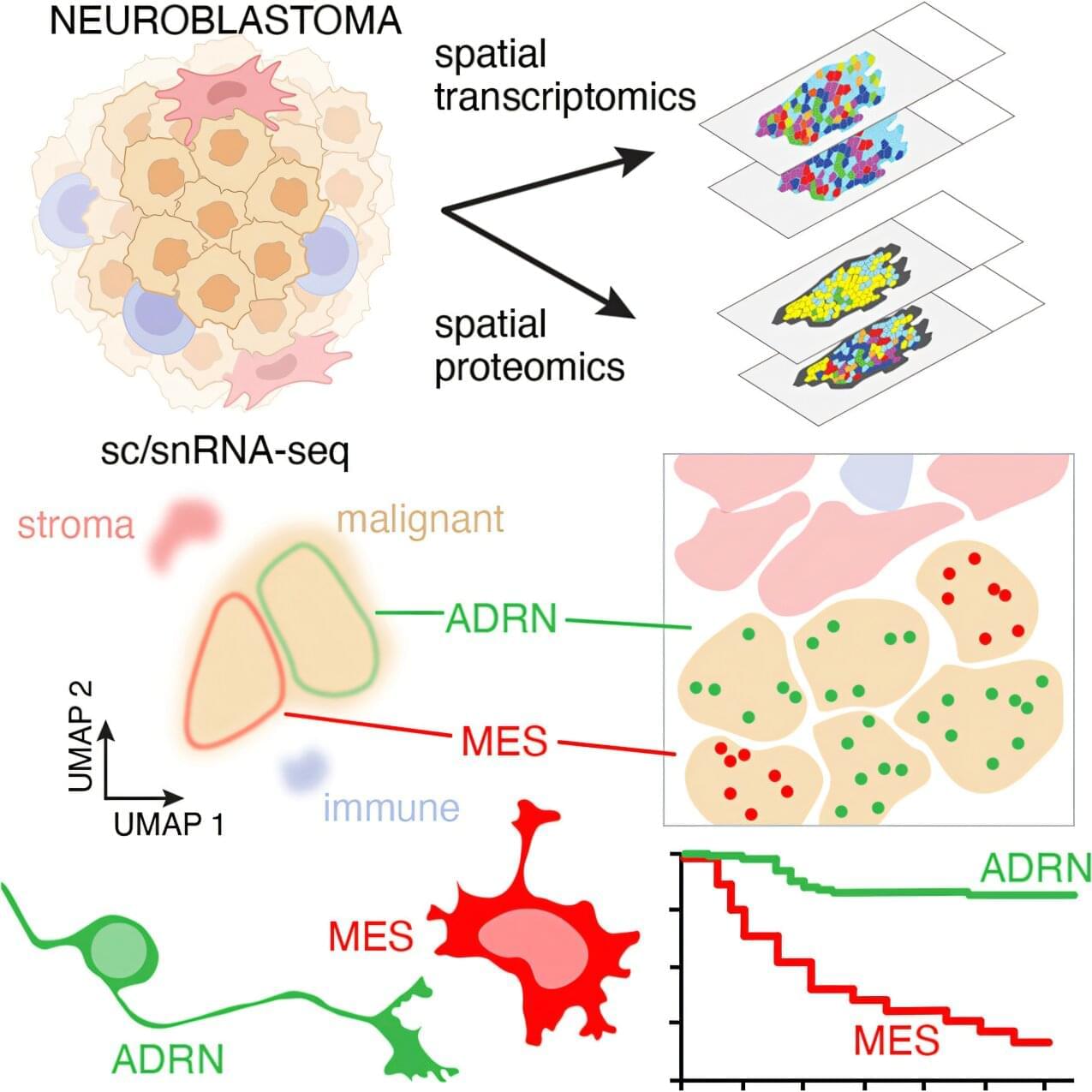
Neuroblastoma is the most common solid tumor found outside the brain in children, yet why some children respond well to treatment while others do not has remained poorly understood. St. Jude Children’s Research Hospital scientists and their collaborators built one of the most comprehensive datasets of neuroblastoma cells to date, using it to define the tumor’s cell types and identify a gene signature marking a malignant cell population tied to poor prognosis.

The team also created patient-derived models that capture this population, which older laboratory cell lines had missed. The findings, published today in Cancer Cell, give researchers a new framework and a set of tools to study high-risk neuroblastoma.

Neuroblastoma cells are divided into two cell states: adrenergic cells (which are associated with low-risk disease) and mesenchymal cells (which are associated with high-risk disease, treatment resistance and poor outcomes). However, directly identifying cancer-related mesenchymal cells in patient tumors has been challenging because the body also contains healthy mesenchymal cells, which can be mistaken for their malignant counterparts, complicating efforts to study them.

Advances in Systemic Treatment of Breast Cancer

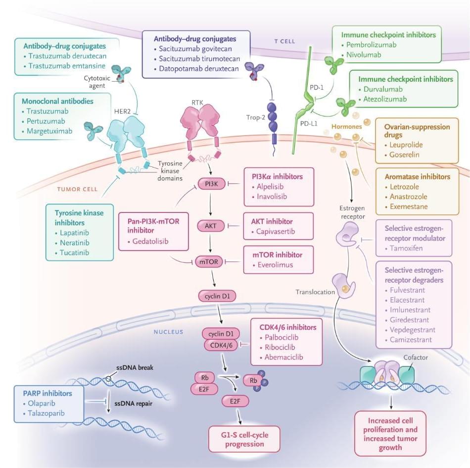
Systemic therapy for breast cancer has evolved rapidly since 2020, driven by the expansion of targeted therapies, antibody–drug conjugates, and immunotherapy across biologic subtypes. The integration of tumor molecular profiling and germline testing has enabled increasingly personalized, biomarker-guided treatment strategies. In hormone-receptor–positive, human epidermal growth factor receptor 2 (HER2)–negative disease, cyclin-dependent kinase 4 and 6 inhibitors remain foundational, whereas phosphatidylinositol 3-kinase pathway inhibitors, oral selective estrogen-receptor degraders, and antibody–drug conjugates have transformed disease management after endocrine resistance. In HER2-positive disease, antibody–drug conjugates — particularly trastuzumab deruxtecan — have improved outcomes and are being used in earlier lines of therapy, a development that is reshaping treatment paradigms.

Light-responsive inks could make tattoos visible on demand, but safety remains unproven

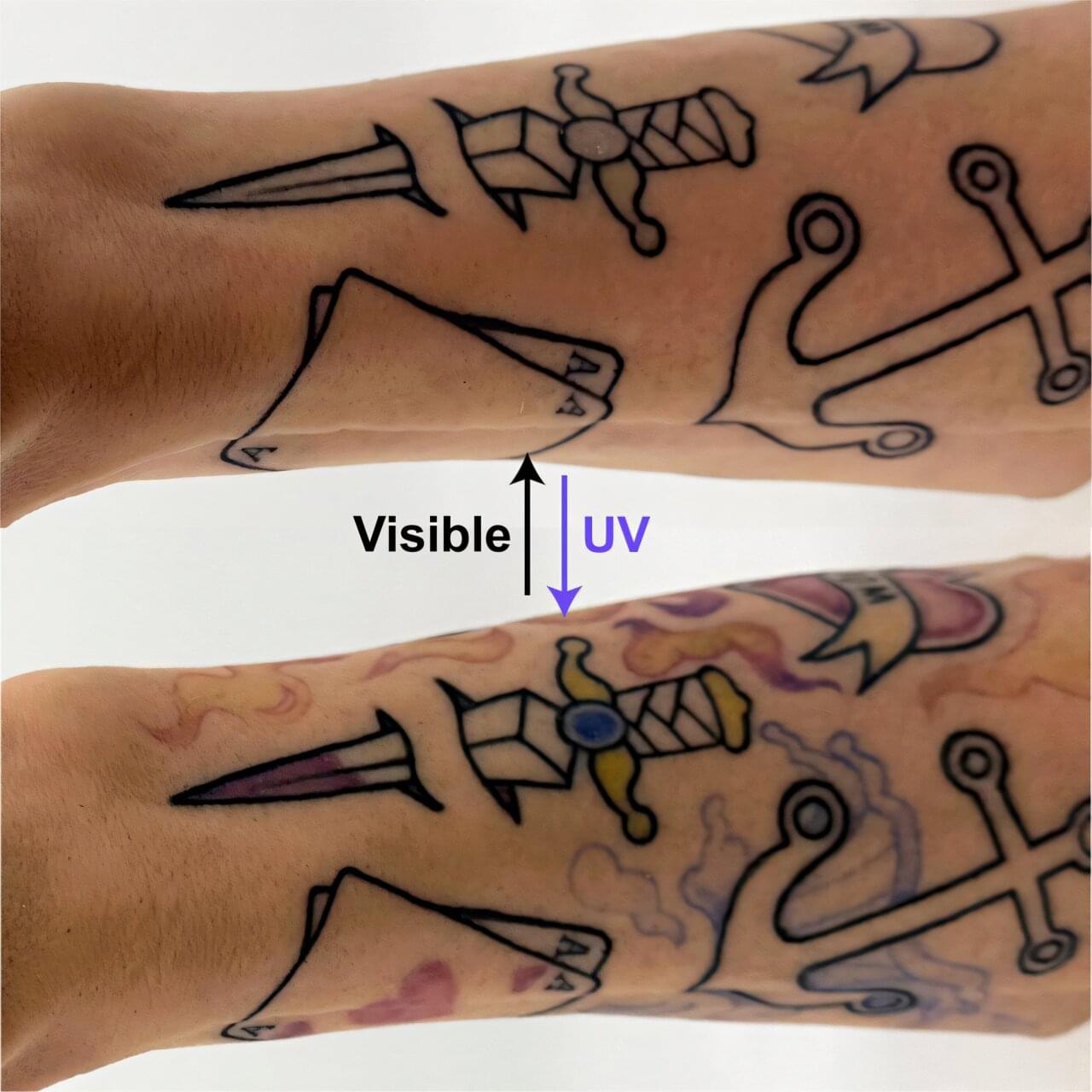
A team of engineers has designed a new line of multicolored tattoo inks that switch on and off with different kinds of light. Described in a study published in Matter & Light, the technology could one day help people hide their tattoos when they have an important business meeting. They could even doodle on their own skin using a simple laser pointer.

“Tattooing is a very ancient technology that remains mostly untapped as a platform for today’s technologies,” says lead author Carson Bruns of the University of Colorado Boulder.

The new inks, known as PhotoTat, are made from dyes and a Plexiglas-like material common in many medical technologies on the market today, including dermal fillers. The research is a passion project for Bruns, who has a lot of his own tattoos, including a green lion on his arm.

New sensing method expands label-free particle detection beyond sensor surfaces

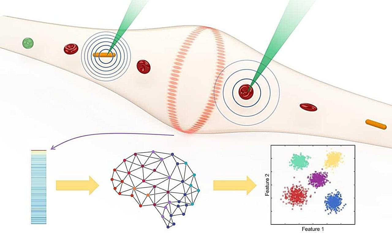
Finding a single cancer cell in a tube of blood is a bit like finding a needle in a haystack. As researchers seek to detect ever-smaller and rarer targets, from viruses to circulating tumor cells in blood, the demand for ultrasensitive, robust sensors that are practical for real-world samples continues to grow.

A major challenge is doing this without adding fluorescent tags or other labels. Most label-free optical sensors work only when the targets pass extremely close to or bind to a tiny sensing area on the device. At low concentrations, many particles never hit that spot, and even when they do, their signals can be drowned out by background noise, limiting detection efficiency and making rare targets especially difficult to find.

This limitation is shared by many optical microsensors, including whispering-gallery-mode (WGM) resonators, among the most sensitive optical sensors ever developed. These devices can detect individual nanoparticles and molecules with exceptional precision, but their sensing region is typically confined to a very small area close to the sensor surface.

Blood cells from 80-year-old donor become stem cells with molecular age under 20

Researchers from the University Hospital of Bonn and the University of Bonn have succeeded in dramatically rejuvenating human cells in a test tube. To do so, they directly reprogrammed red blood cell precursors into stem cells from which neurons can be derived. During this process, the molecular clocks that indicate a cell’s age were reset. For example, blood cells from an 80-year-old were transformed into stem cells with a molecular age of less than 20 years.

Since this rejuvenation process occurred very slowly, it is well-suited for a more detailed investigation of the underlying mechanisms. The results have been published in the journal Aging Cell.

The human body contains hundreds of different cell types. They all originate from the fertilized egg and therefore share the same genetic makeup. However, in the course of their development, they became committed to a specific fate: a skin cell cannot naturally become a liver cell, nor can a blood cell become a nerve cell.