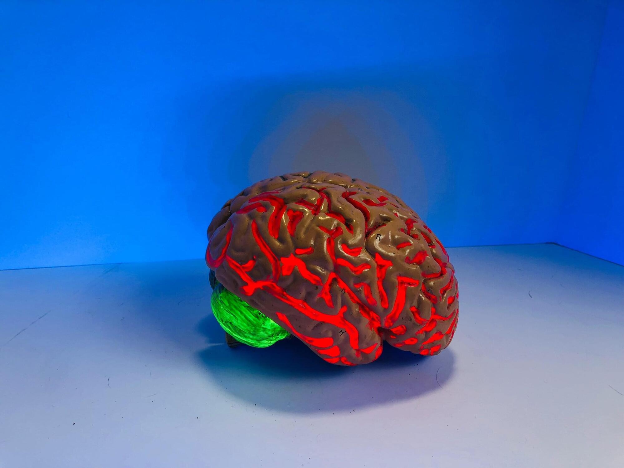
Toggle light / dark theme

Integrative approaches to aging: Mechanisms, antiaging strategies, and emerging biomedical interventions

This imbalance results in dermal thinning, wrinkle formation, and loss of skin elasticity. Both intrinsic aging (chronological) and extrinsic aging (photoaging) contribute to collagen depletion. Studies have shown that UV-induced ROS accelerate collagen breakdown and inhibit new collagen synthesis, exacerbating visible signs of aging. [20]

Collagen is vital for skin firmness and elasticity. Aging, both intrinsic and extrinsic, leads to reduced collagen production and increased enzymatic degradation. Antiaging interventions such as retinoids, marine peptides, and nanoformulations aim to restore collagen levels and improve skin structure.

Understanding these cellular and molecular mechanisms provides the foundation for developing targeted antiaging interventions, ranging from holistic lifestyle modifications to advanced biomedical therapies.

HarmonyGNN boosts graph AI accuracy on four tough benchmarks by up to 9.6%

Researchers have demonstrated a new training technique that significantly improves the accuracy of graph neural networks (GNNs)—AI systems used in applications from drug discovery to weather forecasting. GNNs are AI systems designed to perform tasks where the input data is presented in the form of graphs. Graphs, in this context, refer largely to data structures where data points (called nodes) are connected by lines (called edges). The edges indicate some sort of relationship between the nodes. Edges can be used to connect nodes that are similar (called homophily)—but can also connect nodes that are dissimilar (called heterophily).

For example, in a graph of a neural system there would be edges between nodes representing two neurons that enhance each other, but there would also be edges between nodes that suppress each other.

Because graphs can be used to represent everything from social networks to molecular structure, GNNS are able to capture complex relationships better than many other types of AI systems.

Minimally Invasive Ablation Can Treat Small Kidney Tumors

Among patients with T1a renal cell carcinoma (T1a RCC), ablation and surgical resection showed comparable risks for tumor progression. However, ablation was associated with higher rates of local recurrence but fewer complications and shorter hospital stays than resection or nephrectomy.


“Follow-up data revealed that most local recurrences in patients who underwent ablation were successfully treated with additional ablation or surgery,” the authors wrote.

“[T]his study suggests ablation as a less invasive alternative to surgery for patients with T1a RCC, resulting in a similar high level of oncologic control,” they added.

This study was led by Johanne Ahrenfeldt, PhD, MScEng, Aarhus University Hospital, Denmark. It was published online in Radiology.

APOE4, the Alzheimer’s risk gene, silently undermines bone quality in women

Scientists at the Buck Institute for Research on Aging, along with collaborators at UC San Francisco, have discovered that APOE4, the most common genetic risk factor for Alzheimer’s disease, causes bone quality deficits specifically in female mice, through a mechanism that is invisible to standard imaging and can emerge as early as midlife. The findings, published in Advanced Science, reveal an unexpected biological link between Alzheimer’s risk and skeletal health, and identify a new molecular pathway that could one day inform earlier diagnosis of cognitive decline or guide treatment for bone quality loss in women who carry the APOE4 gene.

“What makes this finding so striking is that bone quality is being compromised at a molecular level that a standard bone scan simply will not catch,” says Buck professor Birgit Schilling, Ph.D., a senior author of the study. “APOE4 is quietly disrupting the very cells responsible for keeping bone strong, and it is doing this specifically in females, which mirrors what we see with Alzheimer’s disease risk.”

Physicians have long observed that people with Alzheimer’s disease suffer bone fractures at higher rates, and that a diagnosis of osteoporosis in women is actually the earliest known predictor of Alzheimer’s. But the underlying mechanism connecting brain and bone health has remained elusive.

New ‘molecular handle’ uses common amino acid to build complex medicines

In a new study published in Nature Communications, a team of chemists has unveiled a radically simple way to attach a highly sought-after “molecular handle,” known as the dichloromethyl group, onto complex compounds. Instead of relying on the aggressive, heavy-metal or radiation-heavy techniques of the past, the team used a common, naturally occurring amino acid called proline to gently choreograph the assembly.

“Rather than forcing these molecules into conventional reactivity modes or circumventing their electronic ambivalence, we harnessed their electronic ambivalence as a design principle,” says Prof. Dmitry Tsvelikhovsky, who led the research team at the Institute for Drug Research at the Hebrew University, alongside Elihay Kuniavsky and Dvora R. Levy.

Compact CRISPR system unlocks targeted in-body gene editing, with up to 90% efficiency

A research team has discovered an enhanced CRISPR gene-editing system that could enable targeted delivery inside the human body—a key step toward broader clinical use. Researchers identified a naturally occurring enzyme, Al3Cas12f, that is small enough to fit into adeno-associated virus vectors, a leading targeted delivery method for gene therapies. They then engineered an enhanced version that dramatically improved gene-editing performance in human cells.

The advance addresses a major limitation in CRISPR technology. Commonly used gene-editing proteins are too large for targeted delivery systems, restricting clinical applications to cells modified outside the body, such as blood and bone marrow.

“Smart delivery of gene editing systems is a powerful notion with broad clinical implications, and this basic science finding takes us a significant step toward that future,” said Erica Brown, Ph.D., acting director of NIH’s National Institute of General Medical Sciences (NIGMS).

Low-frequency wireless sensor tracks artery stiffening in real time with less interference

Wireless sensors used in wearable smart devices and medical equipment must be capable of detecting minute changes while maintaining high operational stability. However, existing technologies often utilize excessively high frequencies, leading to electromagnetic interference (EMI) or potential health risks to the human body. To address these fundamental issues, a Korean research team has developed a low-frequency-based wireless sensor technology.

A joint research team, led by Professor Seungyoung Ahn from the KAIST Cho Chun Shik Graduate School of Mobility and Professor Do Hwan Kim from the Department of Chemical Engineering at Hanyang University, has developed the “WiLECS” (Wireless Ionic-Electronic Coupling System), a low-frequency wireless electrochemical sensing platform that combines ion-based materials with wireless power transfer technology. The research is published in the journal Nature Communications.

Conventional wireless sensors suffer from low capacitance (the ability to store electrical charge), requiring high frequencies in the megahertz (MHz) range to compensate. However, these high-frequency methods can cause tissue heating or signal instability, limiting their practical application in clinical medical settings.

Pain-sensing neurons mapped in unprecedented detail, pointing to new chronic pain drug targets

One in five people worldwide suffers from chronic inflammatory pain. Meanwhile, about two thirds of those affected find little relief from existing pain medications; new therapeutic approaches are urgently needed. “We first must understand precisely how sensory nerve cells trigger pain at the molecular level—in other words, which proteins are involved,” says Professor Gary Lewin, Group Leader of the Molecular Physiology of Somatosensory Perception lab at the Max Delbrück Center in Berlin.

To unravel these molecular processes, Lewin—who has been studying pain for four decades and recently discovered a previously unknown ion channel involved in pain perception—is working closely with systems biologist Dr. Fabian Coscia, Group Leader of the Spatial Proteomics lab at the same center. Coscia co-developed a method called Deep Visual Proteomics that makes it possible to determine the proteome —the complete set of proteins—of specific cells and to create maps detailing the spatial locations of individual proteins.

The researchers combined this technology with electrophysiological methods from Lewin’s group. This enabled them to first identify specific subtypes of pain neurons based on their function and then analyze their protein profiles. The result is a high-resolution molecular map of these nerve cells, which has been published in Nature Communications. The team also demonstrated how the technology can identify potential new drug targets to treat chronic pain.

Designing better membrane proteins by embracing imperfection

Scientists at the VIB–VUB Center for Structural Biology have uncovered a counterintuitive principle that could reshape how membrane proteins are designed from scratch: Sometimes, making a protein less stable helps it fold correctly. In their study published in the Proceedings of the National Academy of Sciences, the researchers demonstrate that introducing carefully placed “imperfections,” a strategy known as negative design, enables synthetic membrane proteins to fold and assemble efficiently in artificial membranes.

Membrane proteins are essential for life and biotechnology, acting as gateways, sensors, and drug targets. Yet designing them from scratch remains notoriously difficult. Unlike soluble proteins, they must navigate a complex folding process while inserting into lipid membranes and during this step, many designs fail.

Traditional protein design focuses on maximizing the stability of the final folded structure. But the new study shows that, for transmembrane β-barrel proteins, this approach can backfire.

These nanotweezers grab thousands of tiny cell packets in seconds and expose their hidden cargo

Justus Ndukaife, associate professor of electrical and computer engineering and Chancellor Faculty Fellow, and his team have developed next generation nanotweezers that better analyze extracellular vesicles and aid in unraveling the mysteries of how cells package molecules and interact with one another. The research was published in Light: Science and Applications journal on March 20, 2026. Graduate student Ikjun Hong helped to perform the experimental characterization under Ndukaife’s direction.

Nanosized extracellular vesicles (EVs), though they vary in size and molecular cargo composition, are an important means for cells to communicate with each other. A significant research opportunity involves analyzing EVs individually to discern their biological roles in diverse diseases as well as leverage them for next generation therapeutics.

Studying single, intact EVs often relies on trapping individual particles, but existing methods face significant limitations. For example, optical tweezers —an approach recognized by the 2018 Nobel Prize in Physics—use a tightly focused laser beam to trap microscopic objects. However, the process is slow, as particles must be captured sequentially, and it is difficult to ensure that a new particle is trapped for each measurement. These constraints severely limit throughput and scalability.

/* */