2 Departments of Medicine and of Pharmacology and Physiology, University of Montreal, Montreal, Quebec, Canada.
3Nephrology Service, Hôpital du Sacré-Cœur de Montréal, Montreal, Quebec, Canada.
2 Departments of Medicine and of Pharmacology and Physiology, University of Montreal, Montreal, Quebec, Canada.
3Nephrology Service, Hôpital du Sacré-Cœur de Montréal, Montreal, Quebec, Canada.
The cell churn that goes on inside our bodies is vast, with hundreds of billions of cells dying off and being replaced every day. Keeping that constant biological overhaul running smoothly is crucial in maintaining good health.
Researchers have now identified a new ‘footprint of death’ that cells leave behind as they die to guide the immune system in its waste-removal role.
It seems this process can also be hijacked by viruses to spread themselves further.
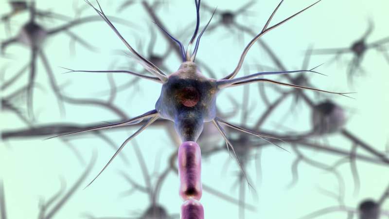
Long before a baby’s ears are functional, the brain is already building the circuitry needed for hearing, according to new research from Johns Hopkins University. Published in the journal Science Advances, the study in mice identifies a previously unknown neural “shortcut” that organizes the auditory system before birth, offering new insight into how the auditory system prepares to process sound and eventually learn language.
While it’s well-known that sound travels from the ear to the auditory cortex, the brain’s hub for hearing, Johns Hopkins researchers discovered a new neural circuit that bypasses the ear entirely. Their findings show that the frontal cortex—the region involved in vocalization—sends signals directly to the auditory cortex, allowing the developing brain to activate hearing-related circuits before external sounds can be heard.
“Our results provide the first direct functional evidence of this biological shortcut that doesn’t go through hearing,” says senior author Patrick Kanold, a professor of biomedical engineering and neuroscience at Johns Hopkins. “It’s a novel brain activity source that can shape the earliest development in mammals, like interpreting information and discerning complex sounds.”
We may age at different rates, but none of us escapes aging. A study in mice and human cells by Stanford Medicine researchers pins much of the blame on a particular type of immune cell’s increasing inability, with advancing age, to gobble up another immune cell type.
So-called tissue-resident macrophages appear to be central coordinators of age-related organ decline. Blocking a single receptor on these cells preserved the youthfulness of multiple organs in mice, including the brain, heart, skeletal and heart muscle, liver, spleen, bone marrow, kidney and colon. The receptor binds specifically to a hormone known to cause inflammation and pain in humans as well as mice.
In mice, selectively disabling this receptor exclusively on tissue-resident macrophages prevented chronic inflammation-driven disorders of aging, including frailty, excessive fat accumulation and heart trouble. It also substantially slowed cognitive decline, said Katrin Andreasson, MD, the Edward F. and Irene Thiel Pimley professor of neurology and neurological sciences.
Researchers at the Icahn School of Medicine at Mount Sinai have identified the immune cell that acts as the architect and coordinator of powerful immune hubs that form inside tumors and plays a key role in antitumor immunity. This discovery could lead to new strategies for making cancer immunotherapies more effective.
The findings, published in Science, reveal that a specialized immune cell called dendritic cell type 1 is essential for building and maintaining structures known as tertiary lymphoid structures (TLSs). These organized clusters of immune cells serve as local command centers, or immune “outposts,” where the body coordinates attacks against cancer directly within tumors.
Previous research has shown that patients whose tumors contain TLSs often live longer and respond better to immunotherapy. Until now, however, scientists did not know what controlled the formation and maintenance of these protective immune hubs.
An international team has succeeded in using a magnetic field to target structures deep within the brain. The researchers injected magnetic nanoplatelets into the relevant region. By doing so, they succeeded in treating movement deficits in mice suffering from Parkinson’s-like symptoms. The new method is less invasive than standard stimulation procedures using implanted electrodes that are currently used to treat certain Parkinson’s disease patients.
The study from Friedrich-Alexander-Universität Erlangen-Nürnberg (FAU), RWTH Aachen and the Universities of Maastricht (the Netherlands) and Leuven (Belgium) has been published in the journal Advanced Science.
In Parkinson’s disease, nerve cells in the brain that produce the neurotransmitter dopamine gradually deteriorate. This affects the motor circuits and leads to tremors and other movement disorders. A brain pacemaker may help some patients. This is a small device that is implanted under the collarbone. From there, it stimulates a region deep within the brain called the subthalamic nucleus (STN for short). This changes pathological activity in these neural circuits and can alleviate movement disorders.
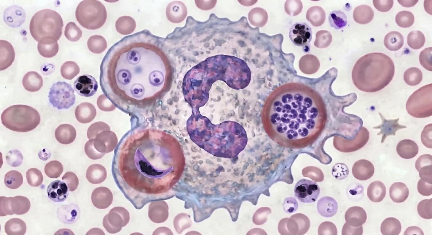
Neutrophils have recently received increased attention in cancer because they contribute to all stages of cancer. Neutrophils are so far considered to have a short half-life. However, a growing body of literature has shown that tumor-associated neutrophils (TANs) acquire a prolonged lifespan. This review discusses recent work surrounding the mechanisms by which neutrophils can persist in the tumor microenvironment (TME). It also highlights different scenarios for therapeutic targeting of protumorigenic neutrophils, supporting the idea that, in tumors, inhibition of neutrophil recruitment is not sufficient because these cells can persist and remain hidden from current interventions. Hence, the elimination of long-lived neutrophils should be pursued to increase the efficacy of standard therapy.
The international guidelines for diagnosing multiple sclerosis (MS), called the McDonald criteria, underwent their most significant overhaul in a decade in 2024. The internationally recognized framework is used to diagnose MS by combining clinical, imaging and laboratory findings. Dr. Jiwon Oh, director of the BARLO MS Centre at St. Michael’s Hospital, was among the experts who helped write them.
Now, in a commentary published in Nature Medicine, Oh has brought together nearly 30 of the world’s leading MS clinicians to critically evaluate what those changes mean and where the field must go next to improve health outcomes for people with MS. Many of its co-authors also helped develop the revised criteria in 2024.
“This commentary looks critically at the new criteria and explains why these changes matter, what challenges may arise as they’re used, and what can be done to address them,” explains Oh. “It also takes a broader look at where the field needs to go next.”
Researchers have demonstrated a novel AI model that can predict which DNA molecules bind with which other DNA molecules. Providing a more thorough understanding of these hypercomplex binding relationships has utility in applications ranging from biomedical diagnostic tools to DNA computing.
“We often think about binding as a very simple relationship – Molecule A binds to Molecule B,” says the co-corresponding author of the study. “But in biological systems, it’s far from simple. Molecule A may bind to dozens of other molecules, to varying degrees.
Capturing that hypercomplexity is a significant challenge, but it is critical if we want to better understand natural genetic systems, says the author. And capturing that hypercomplexity is also critical if we want to develop tools that make full use of biomolecules, such as diagnostic tools that are sensitive to genetic differences or DNA computing systems that rely on DNA to store and retrieve data.