If Spider-Man were real, he could help scientists revolutionize healthcare. Since he’s not, researchers develop creative ways to produce spider silk to study.
Category: biotech/medical – Page 11
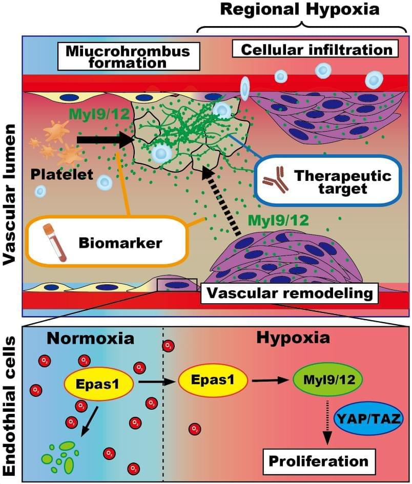
HypoxiaInduced Epas1Myl9/12 Axis Shapes the Pathology of Pulmonary Hypertension
BACKGROUND: Pulmonary hypertension (PH) is a progressive cardiopulmonary disorder characterized by vascular remodeling, abnormal vasoconstriction of small lung arteries, and right heart failure. Hypoxia causes vascular damage, leading to vessel stenosis or occlusion by aberrant endothelial cells, hypertrophy of the tunica media, and thrombus formation. But the precise molecular mechanisms underlying the pathology of PH have been uncertain. METHODS: To investigate the pathogenic role of Myl (myosin light chain) 9/12 in PH, we utilized the Sugen/hypoxia mouse model, generated by administration of the VEGF (vascular endothelial growth factor) receptor inhibitor SU5416 under hypoxic conditions (10% O2). Lung tissues of patients with PH and human lung microvascular endothelial cells were used to examine their endothelial changes.

SingleCell Studies Advance Understanding of the Genetic and Molecular Basis of Atherosclerosis
Foundational models pretrained on millions of single cells (Geneformer, scGPT, and scBERT) now provide transferable embeddings that can help improve and automate cellular annotation, integration, cross-species mapping, and zero-shot predictions.49–51 While there has been considerable contribution to pretraining with immune and tumor data sets, other cell type–specific data for vascular resident parenchymal cells remain sparse, and the applications in atherosclerosis are still emerging. Therefore, overreliance on early foundational models may lead to mislabeling atherosclerosis-specific subtypes and rare cell states, and this area is still being actively investigated with improved consensus in cell types, such as vascular cells (SMC), forthcoming. While promising, we anticipate that these tools will improve with further fine-tuning and robust vascular tissue validation, and interpreted with pathway and genetics-based constraints. Currently, with more modest-sized vascular disease single-cell data sets available, probabilistic variational autoencoder–based methods, such as scVI, MultiVI, and GLUE,44,45,52 offer a good tradeoff between robustness, interpretability, and analytical efficiency. Another exciting area of research is expanding through the development and application of in silico perturbations. While TF perturbation using tools such as CellOracle has provided a first step, improved regulatory network predictions have long been pursued but are still in early stages of implementation.53,54
Deep learning approaches to study TF-DNA interactions are now being utilized to advance our understanding of the DNA regulatory grammar.55 Beyond simple chromatin syntax predictions, deep learning models can provide functional insights, affinity predictions for TF cooperativity, link allelic variation, and chromatin accessibility to cellular epigenetic and transcriptional functions (see below).56 Implementation of a deep learning approach has dramatically extended the capability of scATAC-seq to identify at single basepair resolution the TF motifs that are functional in a cell-specific context to modulate chromatin accessibility, TF binding, and gene expression (Figure 2). Furthermore, allelic variants that are identified with this method are highly enriched among those associated with the complex human traits and diseases that are being investigated. ChromBPNet is a fully convolutional neural network that uncovers the genomic grammar at dynamic enhancers in loci of interest.
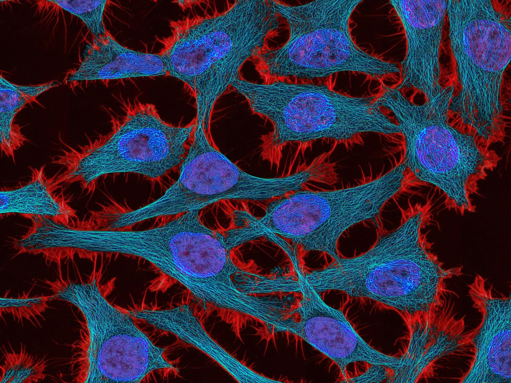
A new drug that can potentially overcome cancer therapy resistance
Researchers at Baylor College of Medicine have developed a drug called CS18 that disrupts cancer cells’ ability to survive therapy. The findings, published in Science Advances, support exploring the possibility of using CS18 to treat human cancer in the future.
“Therapeutic resistance is a main obstacle to achieving effective and durable cancer treatments,” said corresponding author Dr. Weei-Chin Lin, professor of medicine—hematology and oncology—and molecular and cellular biology at Baylor. “While some therapies are effective at the beginning, many patients eventually relapse because cancer cells can activate compensatory and convergent biological pathways that allow them to overcome the toxic effects of therapy, promoting survival.”
In the current study, the researchers’ goal was to develop a drug that would target a “biological switchboard”—topoisomerase IIβ-binding protein 1 (TopBP1)—that controls several cancer-driving pathways at once and to determine whether this strategy could deliver durable responses and overcome resistance.
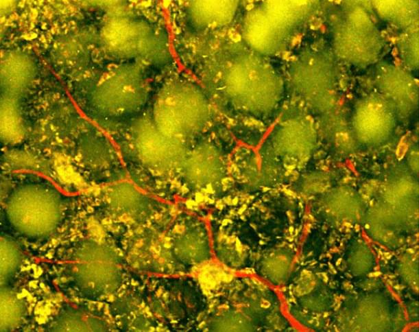
Injectable biomaterial harnesses the immune system to promote brain repair after a stroke
Biomedical engineers at Duke University have developed an injectable biomaterial that helps transform the cavity left behind after a stroke caused by a blood clot into an environment more supportive of repair. By recruiting the body’s own immune cells, the treatment promoted the growth of new blood vessels, supported neural remodeling and improved motor performance in mice. The work appears in Cell Biomaterials.
Every year, millions of people experience strokes caused by blood clots, called ischemic strokes. Immediate treatments, including clot-dissolving drugs and mechanical removal of the clot, can restore blood flow and preserve threatened brain tissue. But these treatments cannot replace tissue that has already been lost.
Large strokes can result in substantial tissue loss and leave behind a cavity at the site of the injury. After the clot is removed, recovery relies largely on rehabilitation, which helps surviving brain circuits adapt but does not directly repair the damaged area.
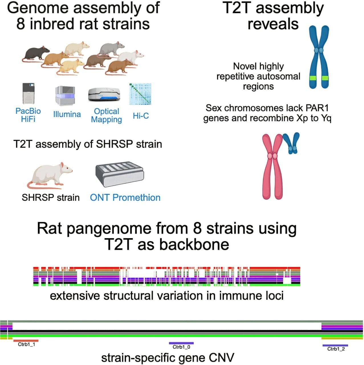
Telomere-to-telomere brown rat genome could sharpen disease research models
Researchers have created the most complete genetic profile of the brown rat to date, according to a UTHealth Houston-led team, paving the way for scientists to more accurately investigate genetic links to conditions like heart disease, kidney disease, high blood pressure and stroke.
The research, published in Cell Genomics, was led by corresponding author Peter Doris, Ph.D., director of the Center for Human Genetics at The Brown Foundation Institute of Molecular Medicine within McGovern Medical School at UTHealth Houston.
The assembly of the brown rat’s genome provides a complete genetic fingerprint and reveals that the brown rat’s DNA is more complex than scientists previously understood. In addition to uncovering more than 60 new genes, many of which were previously difficult to sequence and are thought to play a role in immunity and other biological processes, the team discovered that brown rat sex chromosomes differ significantly from those in humans.
Reversing Cellular Age: The Scientist Who Helped Create iPS Cells Reveals What’s Next
For most of human history, scientists believed that once a cell became a skin cell, a neuron, or a heart cell, that identity was permanent. Then a group of researchers discovered something extraordinary: cells could be reset. My guest today was there when that discovery happened.
Dr. Koji Tanabe, Ph.D. is Founder and CEO of I Peace (https://ipeace.com/en/), one of the world’s leading companies advancing induced pluripotent stem cell — or iPSC — technology from the research laboratory into scalable clinical manufacturing.
Dr. Tanabe occupies a truly unique place in modern biomedical history. He earned his Ph.D. in the laboratory of Nobel Laureate Dr. Shinya Yamanaka at Kyoto University and was the second author on the landmark scientific paper that first demonstrated the successful creation of human induced pluripotent stem cells — a discovery that fundamentally changed regenerative medicine and ultimately earned Dr. Yamanaka the 2012 Nobel Prize.
After helping establish one of the most important technologies in modern biology, Dr. Tanabe continued his work at Stanford University in the laboratory of Dr. Marius Wernig, a pioneer of direct cellular reprogramming, where he investigated how mature blood cells can be directly converted into neurons and explored the molecular mechanisms that govern cellular identity.
In 2015, Dr. Tanabe founded I Peace with an ambitious vision: to make clinical-grade iPS cells accessible at industrial scale. Today, the company has developed automated GMP manufacturing platforms capable of producing personalized and clinical-grade iPS cells for researchers, pharmaceutical companies, and regenerative medicine programs around the world.
On the episode we’ll explore how far the field has come since those first groundbreaking experiments nearly two decades ago, where regenerative medicine stands today, the growing role of iPS cells in drug discovery and transplantation, the excitement surrounding in vivo reprogramming and partial cellular rejuvenation, and what may ultimately become possible when every individual has access to their own personalized stem cell bank.
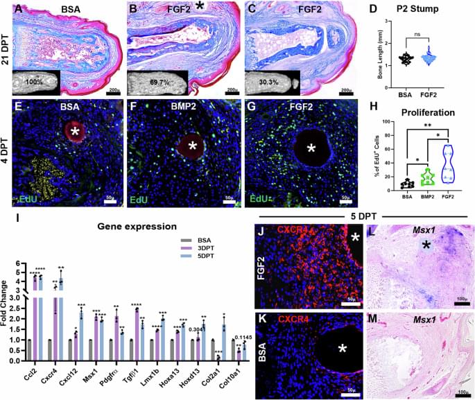
Digit regeneration in mice is stimulated by sequential treatment with FGF2 and BMP2
Basically whole body regeneration is definitely possible we just need to right genetic code to push the regeneration button in the human body much like how these mice had their digits regenerated so too we can regenerate just like Deadpool or even the axolotl.
Wound fibrosis after amputation in mammals is replaced with regeneration of amputated structural elements by sequential FGF2/BMP2 treatment. Regenerated tissues include phalangeal/sesamoid bones, tendon/ligament, synovial joint, articular cartilage.
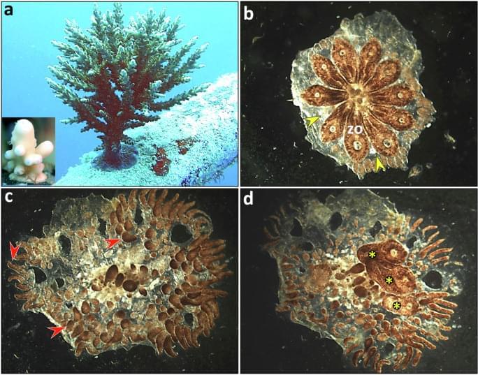
From fragment to form: wholebody regeneration in a model urochordate Medicine
Whole body regeneration is possible just would need to find it in human beings similar genetics.
Rinkevich, Y., Rinkevich, B. From fragment to form: whole-body regeneration in a model urochordate. npj Regen Med 10, 36 (2025). https://doi.org/10.1038/s41536-025-00423-0