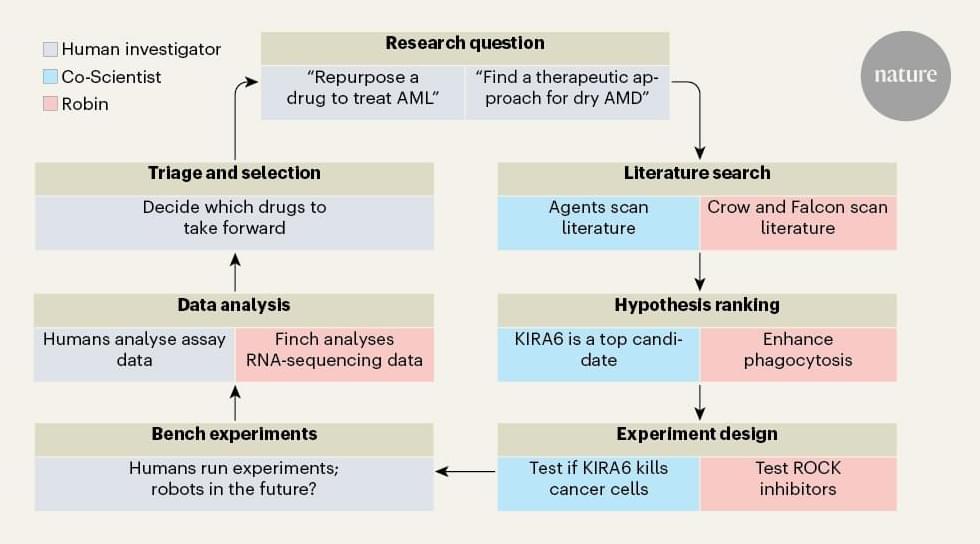
CCR researchers led by Peng Jiang, Ph.D., Senior Investigator in the Cancer Data Science Laboratory, have developed a new tool called the Cancer Immunology

Abstract:
Alzheimer’s disease (AD) is a prevalent neurodegenerative disorder characterized by β-amyloid (Aβ) deposition, tau protein hyperphosphorylation, and synaptic dysfunction. In recent years, 40 Hz sensory stimulation—including visual, auditory, and multimodal modalities—has emerged as a novel, non-invasive intervention demonstrating potential efficacy in both animal models and preliminary clinical studies. Preclinical evidence indicates that such stimulation can markedly reduce cerebral Aβ burden (by approximately 37%–53%), inhibit tau protein phosphorylation, enhance neuronal network synchrony and synaptic plasticity, and improve learning and memory performance. Limited human trials suggest that 40 Hz sensory stimulation is safe and well tolerated in patients with mild cognitive impairment (MCI) and early-stage AD, with a slowing trend in cognitive scale score decline following intervention. This review summarizes the mechanisms of action, experimental evidence from animal models, and advances in clinical application of 40 Hz sensory stimulation in AD prevention and treatment. It further explores the potential for multimodal combination therapies integrating sensory stimulation with cognitive training, pharmacological interventions, and lifestyle modifications, and addresses challenges such as optimal timing of intervention and the influence of ambient electromagnetic fields in real-world settings. Current evidence supports 40 Hz sensory stimulation as a feasible, multi-target, and safe adjunctive intervention; however, its efficacy and applicability must be verified through multicenter, randomized controlled trials with long-term follow-up.

The first human trial of this age-reversal technology is officially underway.
After showing promise in mice and monkeys, researchers are now testing whether the same approach can rejuvenate human cells with the long-term goal of applying it across the entire body.
Chronic stress, the prolonged exposure to psychological and/or physical strain, is known to be a risk factor for depression, anxiety and some other psychiatric disorders. Past studies suggest that chronic stress disrupts the integrity of myelin, a fatty insulating layer that surrounds nerve fibers and helps electrical signals travel efficiently between brain cells.
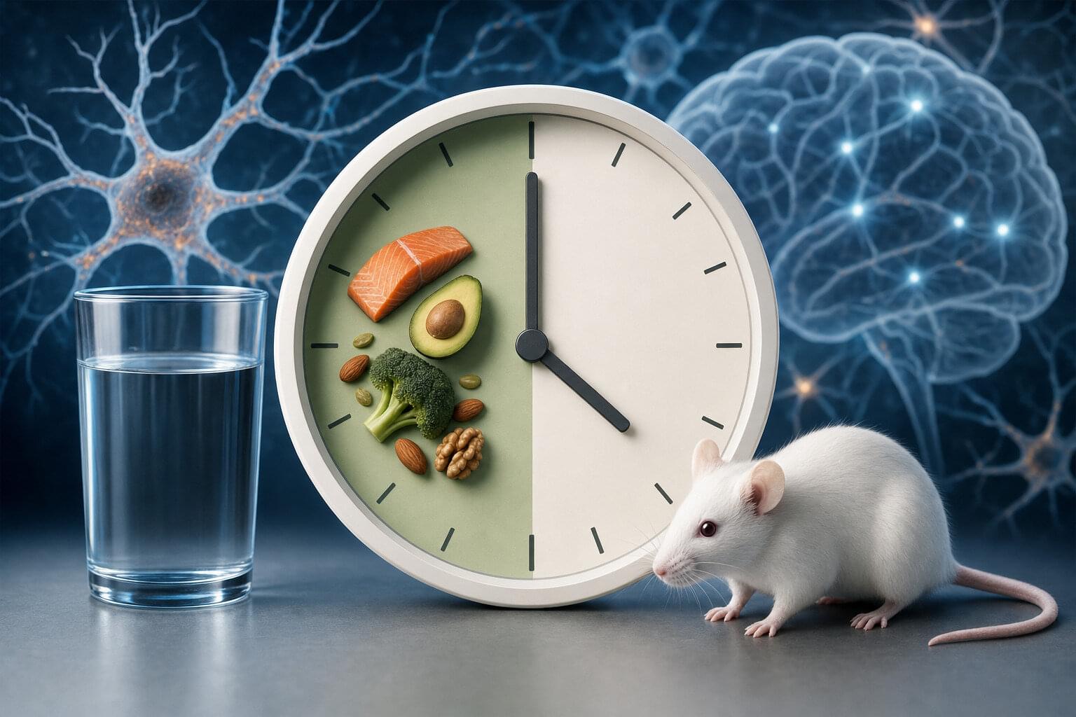
Identifying lifestyle changes that can reverse or diminish the adverse effects of chronic stress on the brain could be advantageous, as they could potentially help prevent or delay the onset of various psychiatric conditions. Recently, some researchers have been exploring the potential brain benefits of intermittent fasting (IF), a dietary pattern that entails alternating between set periods of eating and fasting.
Past findings suggest that IF can improve people’s metabolism and help reduce inflammation, the body’s natural response to disease or injury. Yet its effects on people’s mental health and well-being have not yet been clearly determined.
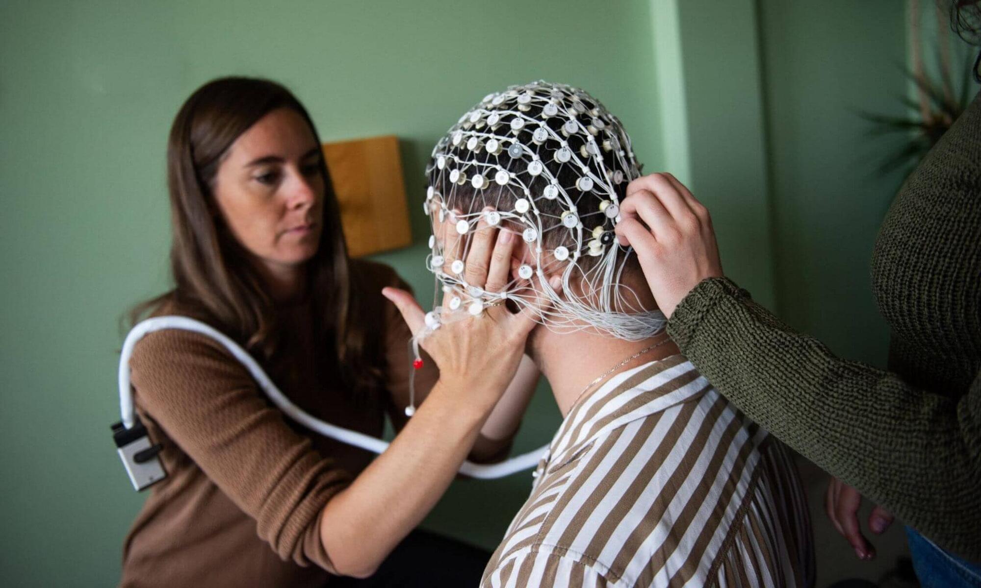
In the brain, specific electrical waves are associated with different states of consciousness. For instance, delta waves—also known as slow waves—are especially prevalent during deep sleep, as well as during states of unconsciousness induced by coma and general anesthesia. They are considered a “signature” of these altered states of consciousness.
Over a decade ago, research showed that it is possible to amplify these delta waves through highly precise auditory stimulation, a technique initially studied in the context of sleep.
Now researchers at Université de Montréal are bringing this technique into the operating room to help optimize general anesthesia, which also induces a state characterized by abundant delta waves.
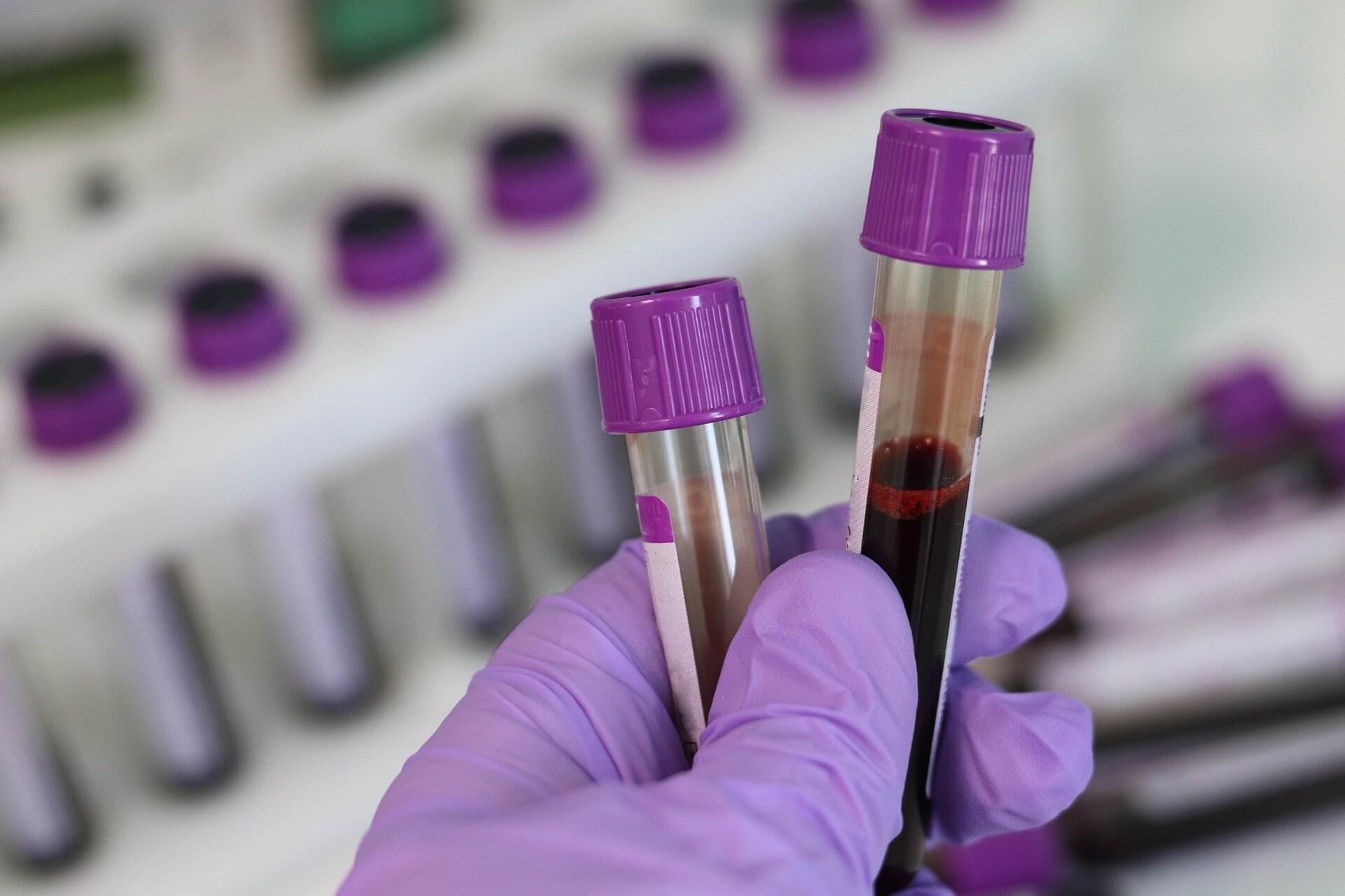
New research, published in The Journal of Immunology, identifies biomarkers of a distinct immune profile that could be used to identify patients at risk for chronic critical illness (CCI) on admission to the intensive care unit (ICU) after traumatic injury. Identifying which patients are at increased risk for CCI could allow doctors to intervene earlier, leading to shorter ICU stays and improved patient outcomes.
“Our findings are highly novel, challenging what scientists have long thought about the immune changes that cause organ dysfunction and mortality in severely injured trauma patients. Rather than the immune system being exhausted, our data show overactivity and dysfunction,” said Dr. Scott Brakenridge, professor of surgery at the University of Washington and senior author of the study.
Severe traumatic injury, such as from a car crash or fall, causes changes to the immune system that can lead to immune and organ dysfunction, as well as recurrent infections. Researchers have long thought this was due to a deficiency in an immune signal, or cytokine, called interferon-gamma (IFN which regulates immune responses.

Address correspondence to: Puneet Opal, Denning Ataxia Center, Department of Neurology, Northwestern Feinberg School of Medicine, Ward 10–332303 East Chicago Avenue, Chicago, Illinois 69,611, USA. Phone: 312.503.4699; Email: [email protected].
A team of researchers at the University of Warwick and Monash University has solved a puzzle that has stumped drug developers for decades: how bacteria naturally create multiple versions of powerful cancer therapies. The breakthrough could accelerate the development of new treatments for hard-to-treat cancers.
Harnessing bacterial enzymes to create drug variants, a strategy known as combinatorial biosynthesis, has long been a goal for scientists. But without understanding how these enzymes interact, progress has stalled.
Published in Nature Communications, the researchers have finally revealed how bacterial enzymes communicate and work together to assemble a family of related anticancer compounds. This family includes romidepsin (Istodax), a clinically approved blood cancer treatment. By understanding this “mix-and-match” process and replicating the principle in the lab, the researchers have established an approach to designing new therapies.