Toggle light / dark theme

Cardiac imaging in oncology: the detection of cardiotoxicity

In this episode of the Heart podcast, Digital Media Editor, Professor James Rudd, is joined by Professor Kazuaki Negishi from Sydney, Australia. They discuss the optimal use of imaging to detect cardiac effects of cancer therapies. If you enjoy the show, please leave us a positive review wherever you get your podcasts. It helps us to reach more people — thanks!

Link to published paper: https://heart.bmj.com/content/111/22/1057.

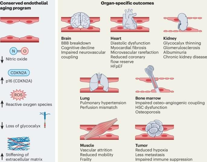
Vascular aging as a driver of organ dysfunction and systemic aging

In this Review, Dimmeler and Augustin discuss microvascular aging as an emerging key driver of tissue dysfunction and systemic decline, highlighting how single-cell and multiomic approaches are uncovering organ-specific vascular vulnerabilities and signaling mechanisms, opening new opportunities for vascular rejuvenation and healthy aging.

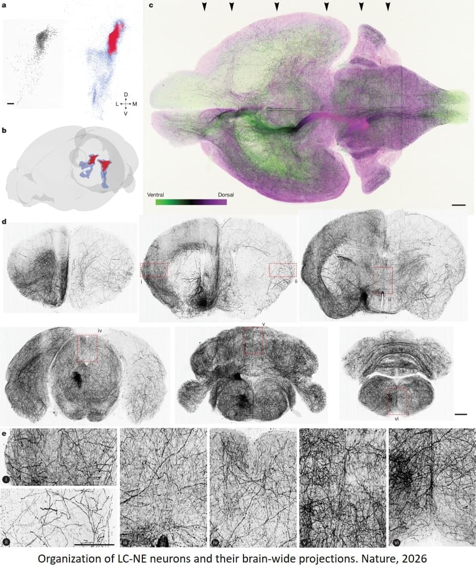
Locus coeruleus noradrenaline neurons mapped!

BACKGROUND: Preeclampsia adversely affects fetal brain angiogenesis, with potential acute and long-term consequences. However, the mechanisms underlying these cerebrovascular alterations remain unclear. We investigated whether TSP-1 (thrombospondin-1), an antiangiogenic glycoprotein, mediates communication between fetoplacental and fetal brain endothelium in preeclampsia. METHODS: Conditioned media from human umbilical vein endothelial cells derived from normal and preeclamptic pregnancies were used to treat human (human cerebral microvascular endothelial cell line) and murine brain endothelial cells. Proteomic analysis of umbilical cord serum identified differentially expressed proteins, and TSP-1 was validated by Western blot. TSP-1 expression was investigated in umbilical endothelial cells (EA.

Utilizing a 3D Spheroid Model and Live-Cell Analysis for Compound Profiling

This application note explores how combining 3D single spheroid models with live-cell analysis can enhance compound screening and drug discovery workflows. Using a library of 880 FDA-approved compounds, researchers evaluated changes in spheroid growth, viability, and morphology over time to gain a more comprehensive understanding of compound activity than traditional endpoint assays can provide. The study demonstrates how live-cell imaging enables continuous, non-invasive monitoring of 3D cell models, delivering rich kinetic data while maintaining cells in a physiologically relevant environment.

The application note highlights the value of phenotypic screening in complex 3D models, allowing researchers to identify cytotoxic, cytostatic, and disruptive compound responses through simultaneous measurement of spheroid size and viability. It also compares findings from 2D monolayer cultures with 3D spheroid assays, revealing important differences in compound sensitivity and response that may improve the translation of in vitro results to in vivo outcomes. By integrating advanced imaging, automated analysis, and 3D cell culture, this workflow provides a powerful approach for identifying promising drug candidates and generating more predictive biological insights early in the drug development process.

Computer-designed protein targets immune receptor linked to inflammation at new ‘undruggable’ site

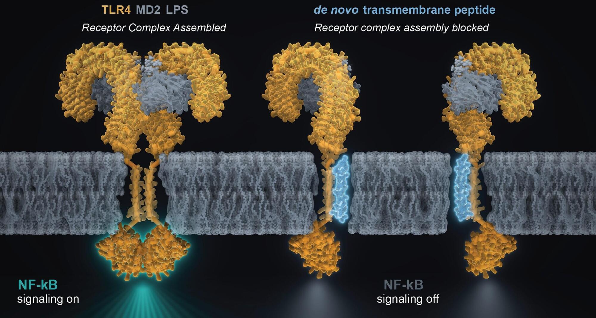
To sense their environment and respond accordingly, cells enlist membrane proteins as communication hubs, receiving molecular messages from outside and triggering responses inside. One of these proteins is Toll-like receptor 4 (TLR4), an immune receptor that plays an essential role in protecting against infections. But overactivity of TLR4 has been linked to inflammatory disorders like sepsis, arthritis and inflammatory bowel disease, making it an attractive target for therapies. Despite its appeal, TLR4 is difficult to manipulate precisely, and no FDA-approved drugs specifically block it.

Now, in a new PNAS study, scientists at Scripps Research engineered a small synthetic protein that can bind to TLR4 within cell membranes and block subsequent inflammatory responses.

The findings advance understanding of what TLR4’s membrane-embedded region does. Rather than a passive anchor, it’s an active determinant of cross-membrane signaling—mechanistic insight that could guide the design of a new class of anti-inflammatory treatments. More broadly, the study outlines new computational tools that other researchers can use to target proteins within membranes.
Smart silicone coatings can change their friction and stickiness as surroundings reshape nanoscale layers

Research led by a group from the University of Newcastle has found a new way to make silicone surfaces—and control how slippery they are. The work was published in the journal Chemistry of Materials.

Most of us have silicone in our homes, especially in the kitchen, where it is often found as a rubbery coating that is long-lasting, water-repellent and, crucially, slippery. Scientists have also wanted to make use of it at the nanoscale, where silicone has significant potential to reduce surface friction for use in medical devices and beyond.

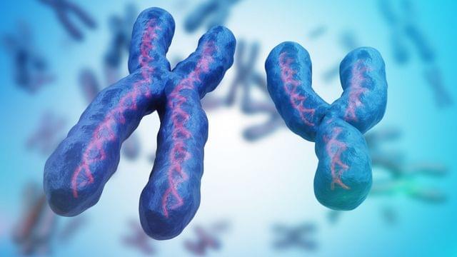
X and Y Chromosomes Play a Direct Role in Aging, Immunity, and Cancer

A new review published in Science brings together growing evidence that the X and Y chromosomes do much more than determining biological sex. They also shape how cells function, age and respond to disease.

Differences in health between men and women have long been attributed mainly to sex hormones such as estrogen and testosterone. However, genes on the X and Y chromosomes also play a direct role, working both on their own and alongside hormones.

The review was co-led by University of Arizona Cancer Center physician-scientist Dr. Dan Theodorescu and Dr. Dena B. Dubal of the University of California, San Francisco. It describes how sex chromosomes influence immunity, metabolism, brain aging, cancer and heart disease, and what this could mean for diagnosis, treatment and the design of clinical trials.

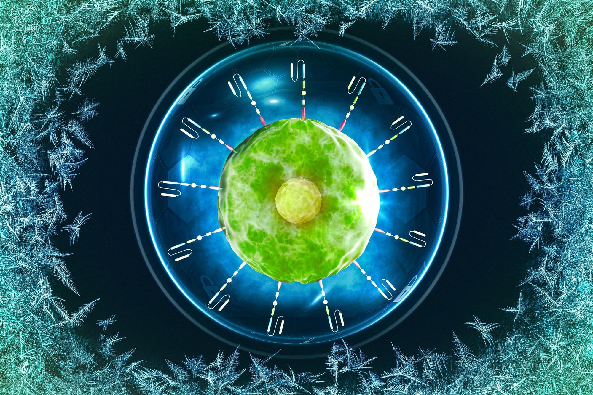
Cell-preservation technique could make CAR-T cell therapy more accessible

Immune cells that are engineered to attack cancer cells, known as CAR-T cells, are used to treat some types of blood cancer. However, only about 5 percent of hospitals in the United States have the ability to generate and deliver CAR-T cells to patients. For many patients, this means the cells need to be frozen and shipped long-distance.

To help make this type of therapy accessible to more people, researchers at MIT have developed a new way to protect the cells from damage that can occur when they are frozen for storage and shipment. Their technique significantly reduces the use of a chemical preservative that is now used to protect the cells, which should make it easier for more hospitals to provide this treatment option to patients.

Instead of treating the cells with a cryoprotective chemical that has to be removed before treatment, the researchers were able to preserve them using a nontoxic antifreeze sugar.

/* */