Myalgic encephalomyelitis (ME) is a chronic, multisystem illness characterized by post-exertional malaise (PEM) and cognitive dysfunction, yet the molecular mechanisms driving these hallmark symptoms remain unclear. This study investigated haptoglobin (Hp) as a potential biomarker of PEM severity and cognitive impairment in ME, with a focus on Hp phenotypes and structural proteoforms.
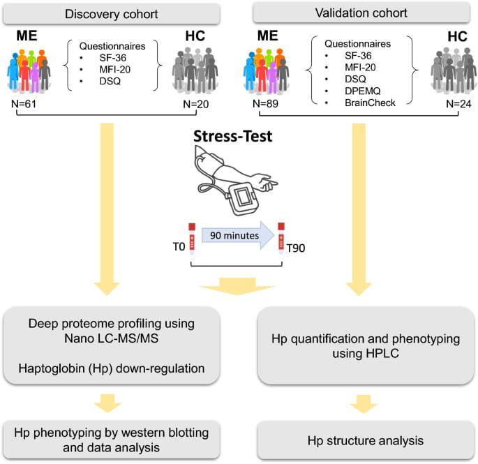
A longitudinal case–control study was conducted in 140 ME patients and 44 matched sedentary healthy controls. In the discovery phase, global plasma proteomic profiling was performed in 61 ME patients and 20 controls before and after a standardized, non-invasive stress protocol in order to induce PEM. Associations between Hp levels, phenotype, and cognitive performance were assessed. In the validation phase, plasma Hp concentrations and proteoform composition were analyzed in an independent cohort of 89 ME patients and 24 controls using high-performance liquid chromatography (HPLC).
ME patients demonstrated a significant reduction in Hp levels following post-exertional stress. Lower baseline Hp concentrations were associated with impaired cognitive performance. Hp phenotypes were differentially associated with symptom burden, with the Hp2-1 phenotype enriched in ME and linked to greater PEM severity and cognitive deficits compared to Hp1-1 and Hp2-2. HPLC analysis revealed altered Hp proteoform profiles in the Hp2-1 subgroup, including increased high-mass tetrameric and pentameric forms and shorter retention times indicative of structural changes. In contrast, the Hp1-1 phenotype was associated with milder symptoms and greater cognitive resilience.