Read this Journal Watch article and more clinical summaries on NEJM Clinician.

Research by the Barcelona Institute for Biomedical Research (IIBB), part of the Spanish National Research Council (CSIC), and the Institut de Recerca Sant Pau (IR Sant Pau) provides some of the first evidence that psychological therapies act as biological stimuli that induce molecular responses measurable through blood biomarkers.
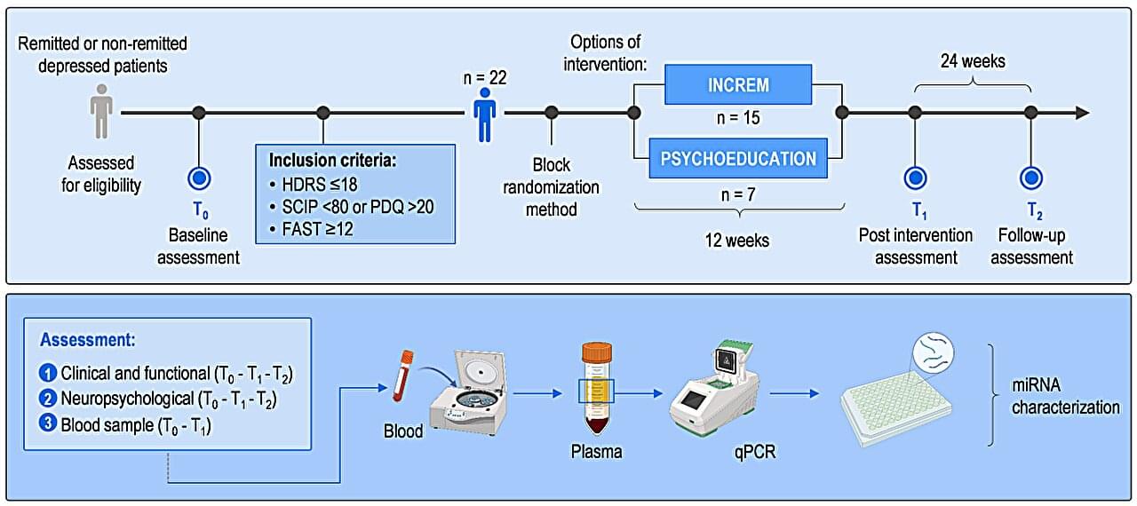
The preliminary study, involving 22 patients with major depressive disorder at Hospital de Sant Pau, reveals that psychotherapy sessions trigger changes in microRNAs—molecules that regulate gene expression in cells—associated with significant improvements in the participants’ cognitive status. The results, published in Scientific Reports, represent an advance toward monitoring patients’ responses to pharmacological treatments and nonpharmacological therapeutic interventions.
The study, led by Dr. Maria J. Portella (IR Sant Pau) and Dr. Analia Bortolozzi (IIBB-CSIC), with Lluís Miquel-Rio (IIBB-CSIC) and Dr. Muriel Vicent-Gil (Hospital de Sant Pau) as first authors, focused on major depressive disorder (MDD). This condition is characterized not only by its effects on mood but also by a broad spectrum of cognitive impairments, including difficulties with attention, memory, processing speed and executive function. These symptoms frequently persist despite treatment and severely affect patients’ quality of life.
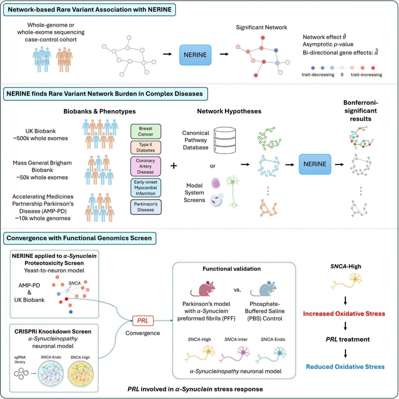
Studies of genetics conducted in yeast cells, human neurons, mice or other model systems often reveal networks of genes that could contribute to complex diseases, such as breast cancer, type 2 diabetes and Parkinson’s disease. But those findings don’t always translate to human biology. Human genetics offers a path to determining which genes among those networks are most relevant to human disease.
Researchers at Harvard Medical School have developed a new statistical framework to link networks identified in models with human genetic data. This could make it faster and easier for researchers to identify which groups of genes are most likely to contribute to a particular human disease, uncover rare disease-causing mutations and zero in on promising therapeutic targets.
The work was published in Cell Genomics.
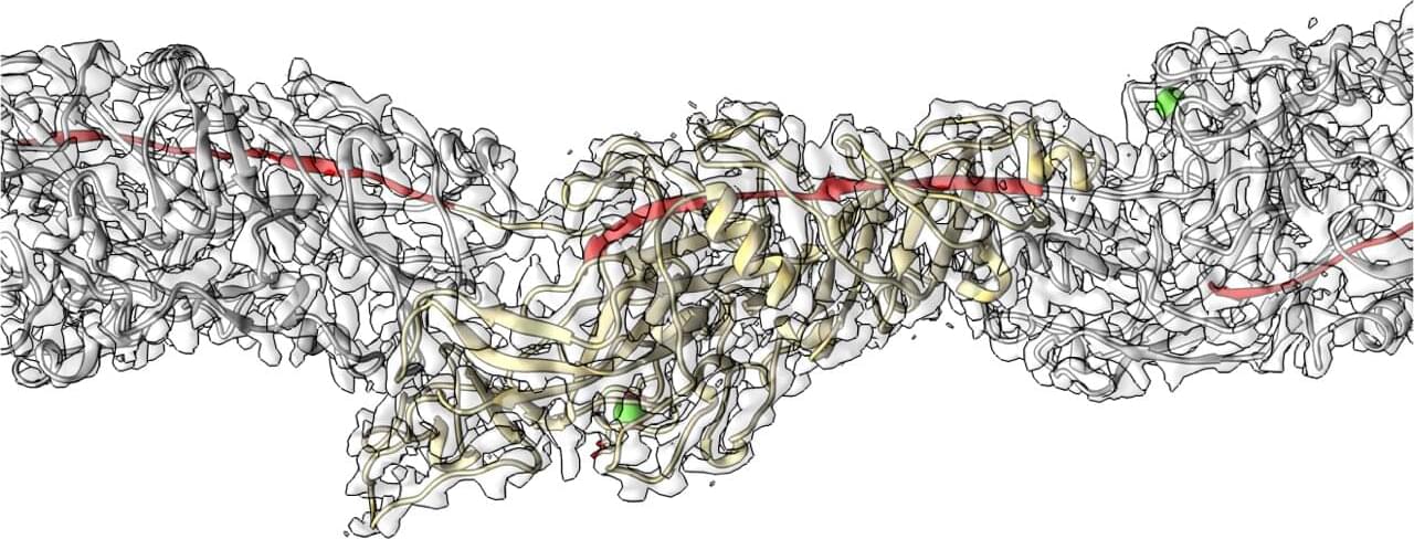
Periodontal (gum) disease is one of the most prevalent diseases worldwide, caused by the bacterium Porphyromonas gingivalis (P. gingivalis). In Japan alone, approximately 80% of adults 30 and older are affected or considered at risk.
Published in Communications Biology, a joint study by the Okinawa Institute of Science and Technology (OIST), Tottori University, Hiroshima University and Nagasaki University provides new insights into how this bacterium causes plaque formation.
Using cryo-electron microscopy (cryo-EM), the researchers reveal the 3D structure of Mfa pili, an arm-like filament that enables the bacteria to stick to host tissues and other microbes.
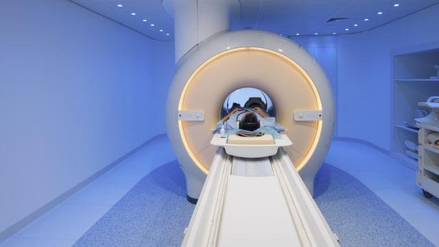
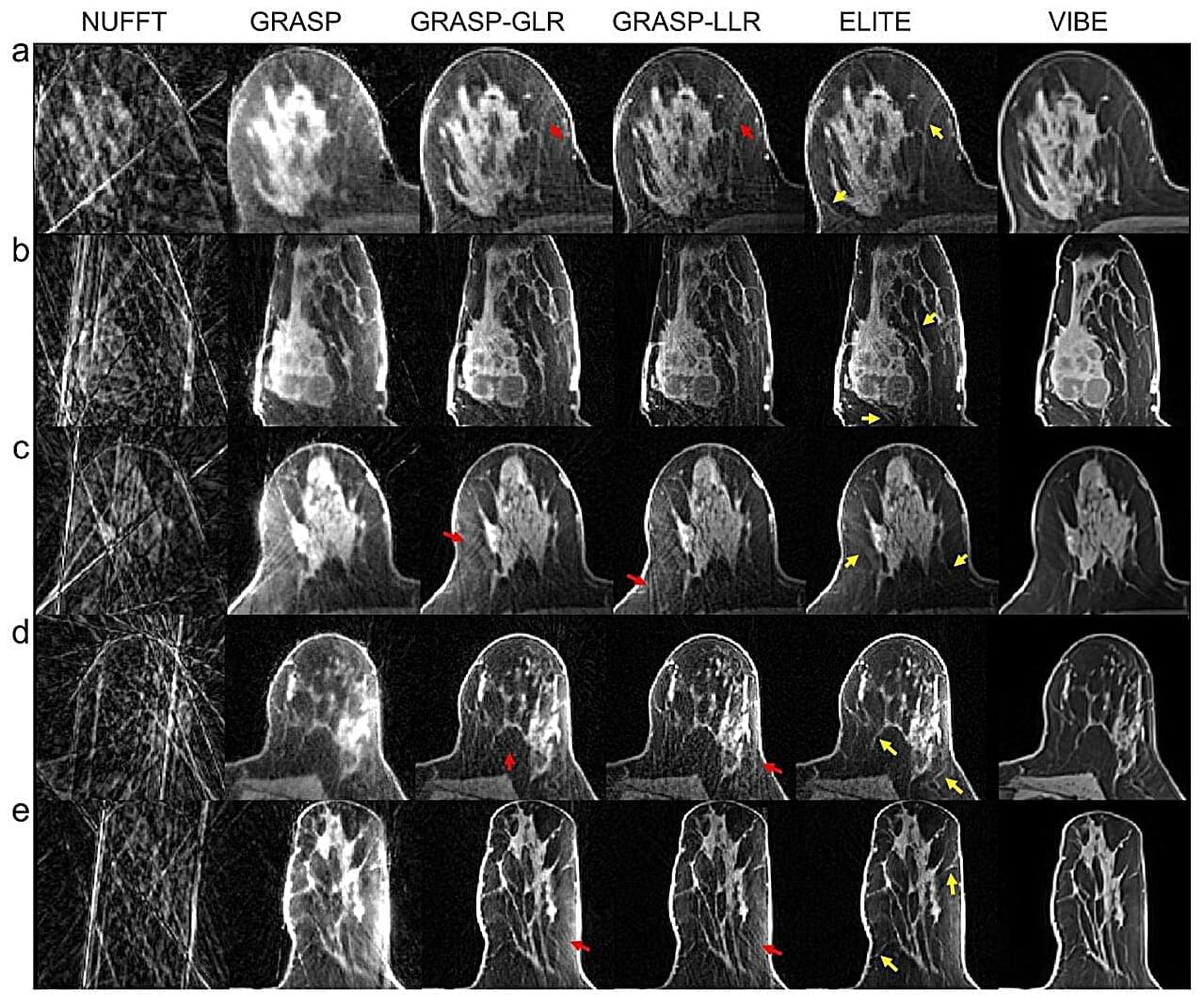
A group of researchers from the Technion and the United States reports a breakthrough in MRI scanning in a paper published in Nature Communications. The researchers developed an innovative method that accelerates and enhances MRI scans for breast cancer imaging, a disease diagnosed in approximately 2.3 million people each year, most of whom are women.
The new method, called ELITE, combines artificial intelligence with advanced mathematical models, enabling dynamic MRI with unprecedented speed and accuracy. This international study brings together expertise in engineering, MRI physics, artificial intelligence and clinical radiology.
Dr. Eddy Solomon of the Technion’s Faculty of Biomedical Engineering, the paper’s lead author, explains that the study focuses on dynamic MRI, a critical technology in breast cancer diagnosis. Dynamic MRI is used primarily for screening populations at high risk for breast cancer and is characterized by exceptionally high sensitivity, with more than 90% accuracy, compared with approximately 50%–60% for ultrasound and mammography combined. However, MRI technology faces a major challenge: Producing highly detailed images usually requires longer scan times, making it difficult to track the flow of contrast material through the examined tissue.
Scientists have discovered a rare genetic condition that causes people to age at a much faster rate, offering fresh insights into the aging process. The study shows for the first time how a “biological clock” present in every cell of the body could contribute to age-related diseases.
Experts say the findings could support the design of future medicines to counter diseases linked to older age, as life expectancies continue to rise across the globe.
The study is published in the journal Nature Genetics.
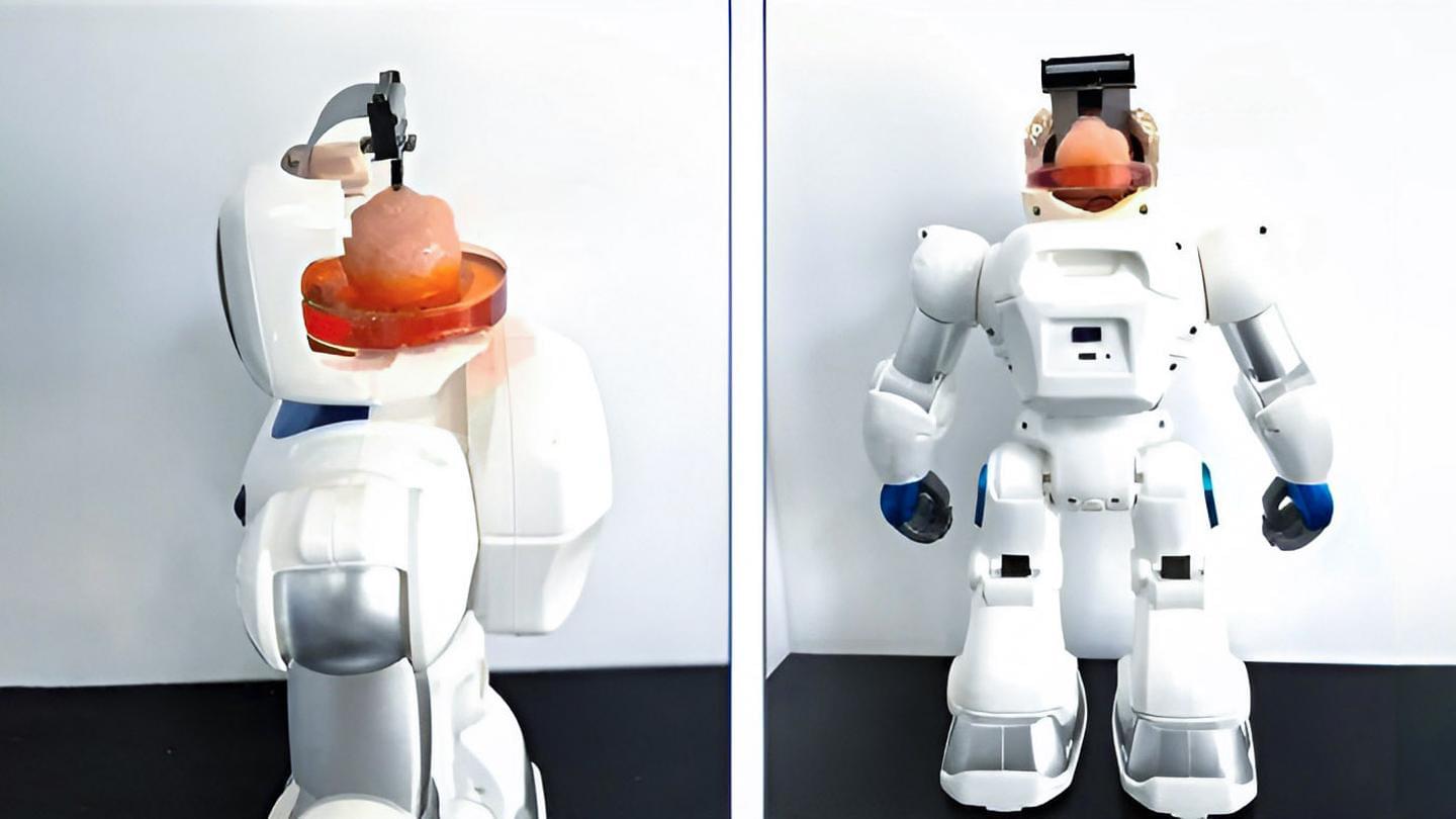
Chinese researchers from Tianjin University and the Southern University of Science and Technology have created a groundbreaking robot powered by a tiny organoid derived from human stem cells grafted to a neural interface. This breakthrough system allows the robot to learn tasks like obstacle avoidance and object manipulation.
Described as the “world’s first open-source brain-on-chip intelligent complex information interaction system,” the technology marks a significant advancement in brain-computer interfaces (BCIs) – devices that translate between neural and computational signals.
The South China Morning Post notes that the scientists grew the organoids from human pluripotent stem cells, which can develop into various cell types, including neural tissue. These synthetic-organic (pardon the oxymoron) brain cells are linked to the robot’s neural interface, enabling communication between the neural tissue and the robot’s systems. Although the presented images of pink brain matter are merely mockups (below), the actual organoids are much smaller.
Destroy them.
Researchers at Tianjin University and the Southern University of Science and Technology in China have created a “human-on-chip” system that combines human brain matter with a neural interface chip and have used the technology to create a hybrid “organoid” robot.
The technology is reported to be an emerging branch of brain-computer interfaces, which aims to combine the brain’s electrical signals with external computing power. The idea behind the technology is to develop brain-like computing.
According to the Global Times, the system uses an artificial brain cultivated in vitro – such as a “brain-like organ” — that can interact with external information through encoding, decoding and stimulus feedback when coupled with electrode chips. In vitro, in this case, means that they’re growing the brain-like organ in a controlled laboratory environment using stem cell technology.
Now, a new study conducted by researchers at Newcastle University and Technische Universität Dresden has used a new lithium MRI technique to reveal that brain lithium levels closely track blood concentrations throughout the day.
Understanding lithium tracking gaps in bipolar disorder
Bipolar disorder affects ~40 million people globally. The mental health condition is characterized by severe shifts in mood, energy, and activity levels. Patients navigate intense emotional states that alternate between mania and deep depression.
Researchers report encouraging early results from a first-in-human clinical trial led by Children’s National Hospital using a new T-cell immunotherapy for children and young adults with some of the deadliest brain tumors, including diffuse intrinsic pontine glioma (DIPG) and relapsed central nervous system (CNS) tumors. These findings, published in Nature Medicine, are particularly significant given the challenges of treating pediatric brain tumors, which remain the leading cause of cancer-related deaths in children. Immunotherapies have been shown to work in blood cancers but rarely succeed in solid tumors, especially brain tumors.
“This study represents an important step toward developing safer and more effective T-cell therapies for children with devastating brain cancers,” said Catherine Bollard, MBChB, MD, senior vice president and chief research officer at Children’s National, and co-senior author of the study. “Even in this early-stage trial focused on safety, we were encouraged to see lasting clinical benefit in several patients who otherwise had very few options.”