5Department of Inflammation and Immunity, Cleveland Clinic Research, and.
6Department of Gastroenterology, Hepatology and Nutrition, Digestive Disease Institute, Cleveland Clinic, Cleveland, Ohio, USA.
The researchers then tested BA-101 together with temozolomide and found that the combination was more effective than either treatment alone. In experiments using mouse models, the combined therapy significantly reduced tumor growth, suggesting that targeting the cancer cells’ resistance mechanism could make existing chemotherapy more powerful.
“Temozolomide resistance remains one of the biggest obstacles in treating glioblastoma,” said Amal. “Our findings suggest that targeting nitrosative stress can restore the tumor’s sensitivity to treatment. While additional studies are needed before this approach can reach patients, these results open an exciting new direction for developing more effective therapies against one of the deadliest cancers.”
The researchers said their findings could point to a new approach in cancer treatment: instead of replacing existing drugs, future therapies could focus on blocking the mechanisms that allow tumors to resist them. If further studies confirm the findings, disabling these survival pathways could allow treatments that have become less effective to regain their ability to attack cancer cells.

A second prostate-specific membrane antigen (PSMA) PET scan changed treatment plans for nearly half of patients whose first scan was negative, according to new research published in the July issue of The Journal of Nuclear Medicine. Findings from the repeat PSMA scans, which included both local and distant disease, resulted in a change in management for nearly 50% of these patients.
Managing recurrent prostate cancer after first-line treatment, such as prostatectomy or radiation therapy, remains a clinical challenge. Although PSMA PET imaging has improved disease detection, 30% of patients still have no detectable disease on initial imaging, even as rising prostate-specific antigen (PSA) levels suggest recurrence. Few studies have examined whether repeating PSMA PET in this situation is worthwhile.
“There is little information on the utility of repeating a PSMA PET after an initial negative scan,” said Ur Metser, BSc, MD, FRCPC, professor of radiology at the University of Toronto and head of the Division of Molecular Imaging at the Joint Department of Medical Imaging at Princess Margaret Cancer Centre in Toronto. “In our study, my colleagues and I sought to determine the benefit of a second PSMA PET scan, as well as to assess predictors for positive PSMA PET scans.”

The 13th Aging Research & Drug Discovery (ARDD) Meeting, the world’s largest conference dedicated to longevity biotechnology, will take place from October 1–3, 2026, at the David Rubenstein Treehouse at Harvard University. Marking the high-profile launch of Boston Longevity Week, this landmark event is officially organized by Insilico Medicine, which also anchors the conference as a Tier 1 sponsor alongside Eli Lilly.
As longevity science rapidly transitions from theoretical concepts to multi-billion-dollar therapeutic pipelines, ARDD 2026 stands as the premier global nexus connecting basic science, clinical research, big pharma, and institutional investors. Moving the conference to Boston-the global epicenter of biomedical innovation-reflects the field’s evolution into mainstream medicine.
Building on the massive momentum of previous years-including ARDD 2025 in Copenhagen, where leadership from Eli Lilly and Novo Nordisk discussed the profound longevity potential of GLP-1 receptor agonists in Nature Biotechnology-the 2026 conference solidifies aging research as a core pillar of healthcare. Top-tier pharmaceutical companies are now actively developing commercial programs targeting fibrosis, immunology, CNS, cardiometabolic diseases, anti-muscle wasting, and cellular rejuvenation.
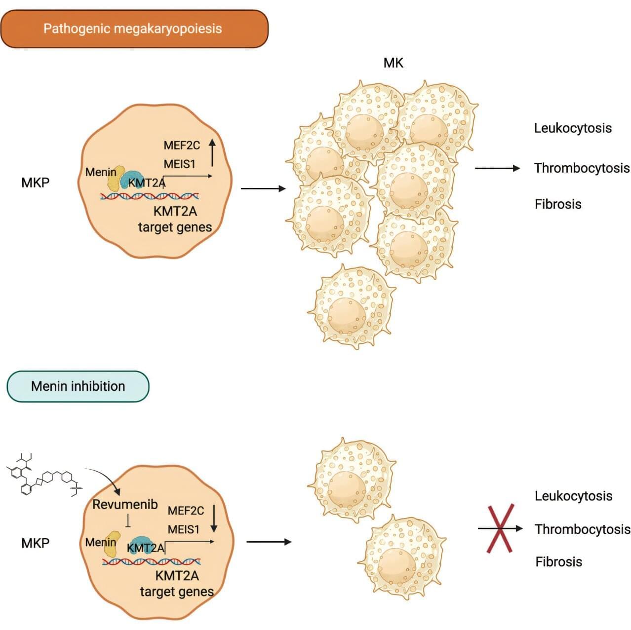
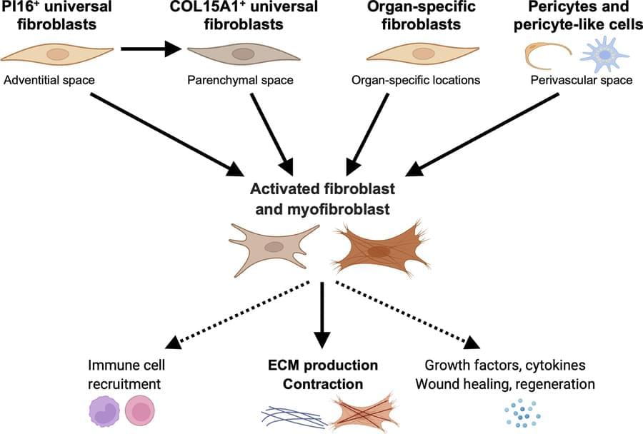
Inhibiting menin, a protein that supports leukemia growth and is already targeted to treat some forms of leukemia, also holds promise for treating myeloproliferative neoplasms. A new study from scientists at St. Jude Children’s Research Hospital showed that inhibiting menin significantly extended survival and reversed multiple disease features in preclinical models. The findings were published today in Cancer Cell.
Menin is best known as a therapeutic vulnerability in certain types of acute leukemia, including those with KMT2A gene rearrangements or NPM1 mutations. Menin inhibitors, such as revumenib, have greatly improved treatment for these cancers and are approved by the Food and Drug Administration (FDA). However, menin inhibition can reduce megakaryocytes (normal platelet-forming cells) and decrease platelet counts. Producing too many megakaryocytes is a hallmark of diseases called myeloproliferative neoplasms, which are slow-developing, rare blood cancers.
John Crispino, Ph.D., MBA, St. Jude Division of Experimental Hematology director and Department of Hematology member, tested whether inhibiting menin could be a viable therapeutic strategy for myeloproliferative neoplasms.
Join us on Patreon! / michaellustgartenphd.
Discount Links/Affiliates:
Blood testing (where I get the majority of my labs, for those who blood test with Quest): https://www.ultalabtests.com/partners… those who blood test with LabCorp: https://www.anrdoezrs.net/click-10161… At-Home Metabolomics: https://www.iollo.com?ref=michael-lus… Use Code: CONQUERAGING At Checkout Clearly Filtered Water Filter: https://get.aspr.app/SHoPY Epigenetic, Telomere Testing: https://trudiagnostic.com/?irclickid=… Use Code: CONQUERAGING NAD+ Quantification: https://www.jinfiniti.com/intracellul… Use Code: ConquerAging At Checkout Oral Microbiome: https://www.bristlehealth.com/?ref=mi… Enter Code: ConquerAging SiphoxHealth Blood Testing (ApoB, GrimAge): https://siphoxhealth.com/mlustgarten Green Tea: https://www.ochaandco.com/?ref=fqbtflod Use Code: ML10OFF Diet Tracking: https://shareasale.com/r.cfm?b=139013… If you’d like to support the channel, you can do that with the website, Buy Me A Coffee: https://www.buymeacoffee.com/mlhnrca Conquer Aging Or Die Trying Merch! https://my-store-d4e7df.creator-sprin…
Blood Testing Essentials (Biological Age, CVD-Risk, Kidney Health and Function):
PhenoAge (Biological Age): https://www.ultalabtests.com/partners…
Measure the Bortz biological clock biomarkers: https://www.ultalabtests.com/partners…
Calculate your biological age using the Bortz clock: https://www.longevity-tools.com/human…

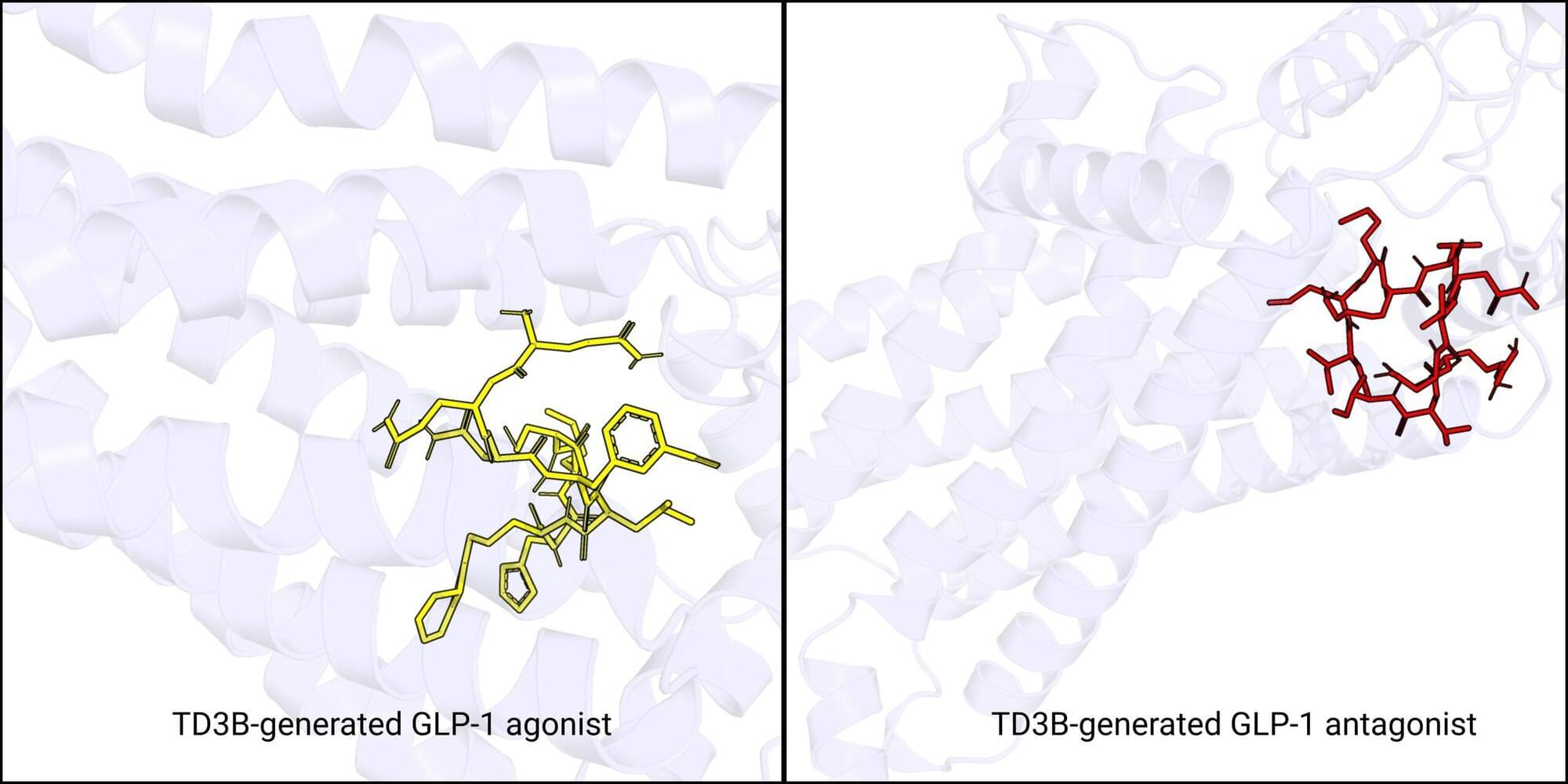
To develop new and better peptides, the short amino acid strings behind medicines like GLP-1 drugs, researchers have used AI to generate candidates and to predict their properties.
However, merging these capabilities into a system that generates peptides likely to activate or block specific targets has proven difficult. In part, this is due to the vast number of possible peptides, but also because predicting how readily a peptide will bind to a target—like G protein-coupled receptors (GPCRs), a family of cell-surface proteins targeted by about one-third of approved drugs—is easier than simultaneously forecasting what effect that binding will have.
Now, researchers at the University of Pennsylvania and The Chinese University of Hong Kong have created TD3B, an AI framework that guides peptide generation toward candidates predicted to have a desired effect. The results, which focus on GPCRs, are described in a paper presented as a Spotlight at the 2026 International Conference on Machine Learning.

Almost everyone will deal with back pain at some point in their lives. Most recover quickly—but for about 20% of people, acute pain becomes a chronic condition that interferes with daily life and keeps them out of the workforce.
Low back pain is one of the leading causes of disability worldwide, and more money is spent managing it in the United States than any other health condition. Despite that, the most effective way to prevent a short-term episode from becoming a long-term problem has not been clear—especially for people who are most at risk.
“Chronic low back pain prevention is a public health issue,” said Michael Schneider, D.C., Ph.D., professor in the School of Health and Rehabilitation Sciences at the University of Pittsburgh and co-principal investigator of the Pitt arm of the study. “The 20% of patients who turn chronic account for 80% of the costs and the suffering. This paper shows that helping people self-manage their pain through a properly trained physical therapist or chiropractor is a great way to mitigate this public health problem.”
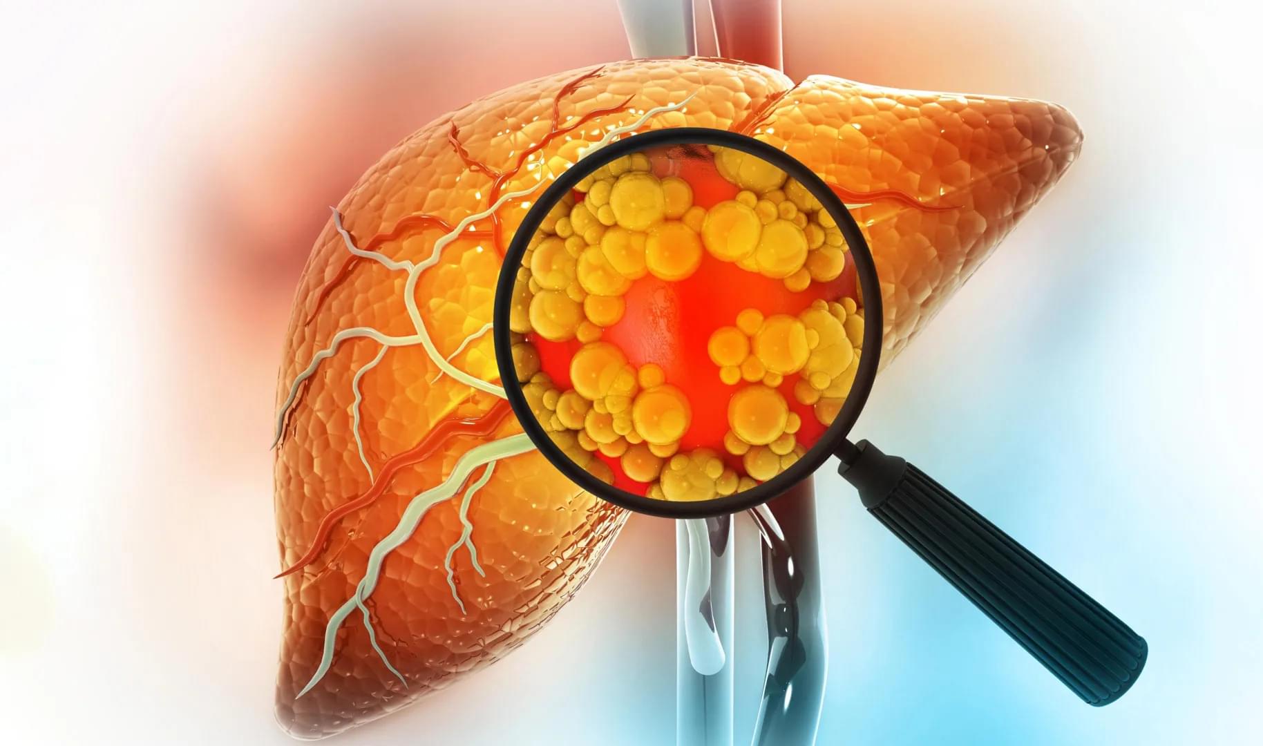
An experimental drug developed at Michigan Medicine has shown the ability to reverse severe fatty liver disease in animal studies by restoring gut health. The findings, published in The Journal of Clinical Investigation, suggest that targeting the connection between the gut and liver could offer a promising new approach for treating metabolic dysfunction-associated steatohepatitis (MASH).
MASH is a serious form of fatty liver disease that affects about 7% of people worldwide. It can progress to cirrhosis, liver cancer, and liver failure, yet effective treatment options remain limited.
The investigational compound, known as DT-109, is a glycine-based tripeptide. Researchers found that it reversed MASH in animal models by interrupting a harmful biological process linking the gut and liver.