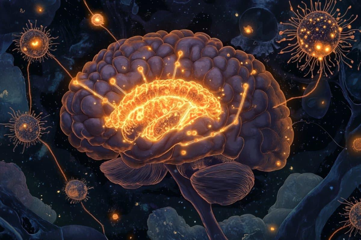
A new study suggests that SSRIs, commonly prescribed antidepressants, may reduce biological markers of Alzheimer’s disease.
Category: neuroscience – Page 391

Study Suggests Key Link That Could Help Explain Autism Development
The importance of our gut microbiota to our health has become increasingly clear in recent years as more and more studies have been released.

Protein misfolding and aggregation in Alzheimer’s disease and Type 2 Diabetes Mellitus
In a diseased condition, most of the time, target proteins attain toxicity following their transition from a α-helix to a β-sheet form [18]. Although numerous functional native proteins possess β-sheet conformations within them, the transition from an α-helix to a β-sheet is characteristic of amyloid deposits [19], and often associated with the change of a physiological function to a pathological one. Such abnormal conformational transition exposes hydrophobic amino acid residues and promotes protein aggregation [18, 20]. The toxic proteins often interact with other native proteins and may catalyze their transition into a toxic sate, and hence they are called infective conformations [18]. The newly formed toxic proteins can repeat this cycle to intiate a self-sustaining loop; thereby amplifying the toxicity to generate a catastrophic effect, beyond homeostatic reparative mechanisms, to eventually impair cellular function or induce cellular demise [21].
Proteins function properly when their constituent amino acids fold correctly [22]. On the other hand, misfolded proteins assemble into insoluble aggregates with other proteins and can be toxic for the cells [18, 20]. Ataxin-1 is highly prone to misfolding due to inherited gene defects that cause neurodegenerative diseases (NDDs), which is mainly due the repetition of glutamine within its amino acid chain; the toxicity of this protein being directly proportional to the number of glutamines [23]. There are 21 proteins that mainly interact with ataxin-1 and influence its folding or misfolding, 12 of which increase the toxicity of ataxin-1 for nerve cells, while 9 of the identified proteins reduce its toxicity [23]. Ataxin-1 resembles a double twisted spiral or helix and has a special structure, termed a “coiled coil domain”, that promotes aggregation. Proteins which possess “coiled coil domain” and interact with ataxin-1 have been reported to enhance promotion of ataxin-1 aggregation and toxic effects [24].
The gradual accumulation of misfolded proteins in the absence of their appropriate clearance can cause amyloid disease, the most prevalent one being AD. Parkinson’s disease and Huntington’s disease have similar amyloid origins [25]. These diseases can be sporadic or familial and their incidence increases dramatically with age. The mechanistic explanation for this correlation is that as we age (and are subjected to increasing numbers of mutations and/or oxidative stress causing changes to protein structure, etc.), the delicate balance of the synthesis, folding, and degradation of proteins is disturbed, ensuing in the production, accumulation and aggregation of misfolded proteins [26].
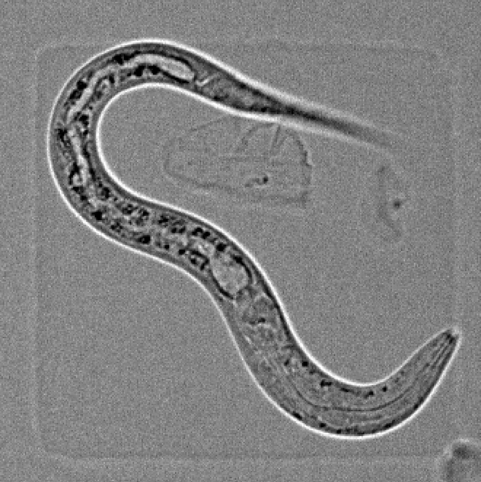
The sleep switch: How one brain signal turns sleep on and off
People spend about a third of their lives asleep. Yet, surprisingly little is known about how our brains control falling asleep and waking up. Now, researchers led by Prof. Henrik Bringmann at the Biotechnology Center (BIOTEC) of TUD Dresden University of Technology discovered another piece of this puzzle. The team showed that a single brain signal acts like a biological switch—both triggering sleep and ending it.
Their findings, published in the journal Current Biology, were made possible by studying a tiny roundworm, C. elegans, a powerful model organism in biology.
“It is really important to be able to fall asleep, but just as important to wake up too,” says Prof. Bringmann, research group leader at BIOTEC who led the study.
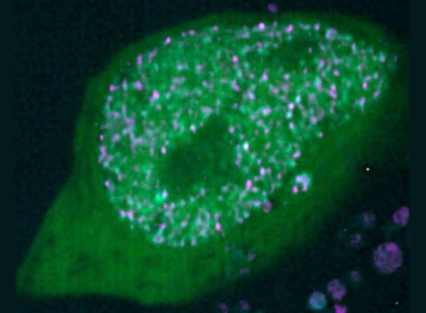
Olfactory neurons use unexpected ‘solid’ clusters to achieve genetic precision
A new study published in Nature reveals how olfactory sensory neurons (OSNs) achieve extraordinary precision in selecting which genes to express.
The mechanism is surprising in that it involves solid-like molecular condensates that last for days, helping to solve a long-standing puzzle in genome organization.
The research, led by Prof. Stavros Lomvardas from Columbia University, addresses one of biology’s most intriguing questions: How do olfactory sensory neurons in the nose manage to express only one olfactory receptor (OR) gene out of approximately 1,000 available options?
Decoding Brain Aging: A 10-Year Study Opens Data for Discovery
A decade-long brain health study has released its full dataset, offering rare longitudinal insights into how cognition and brain structure change across adulthood.
New study identifies gateway to conscious perception
Researchers investigating the thalamus and its role in consciousness believe the area acts as a ‘gate’ to stimuli that reach, or don’t reach, conscious perception.
Can you upload a human mind into a computer? Here’s what a neuroscientist has to say about it
Science has done many things that seem miraculous. Why not transfer your consciousness to a machine?
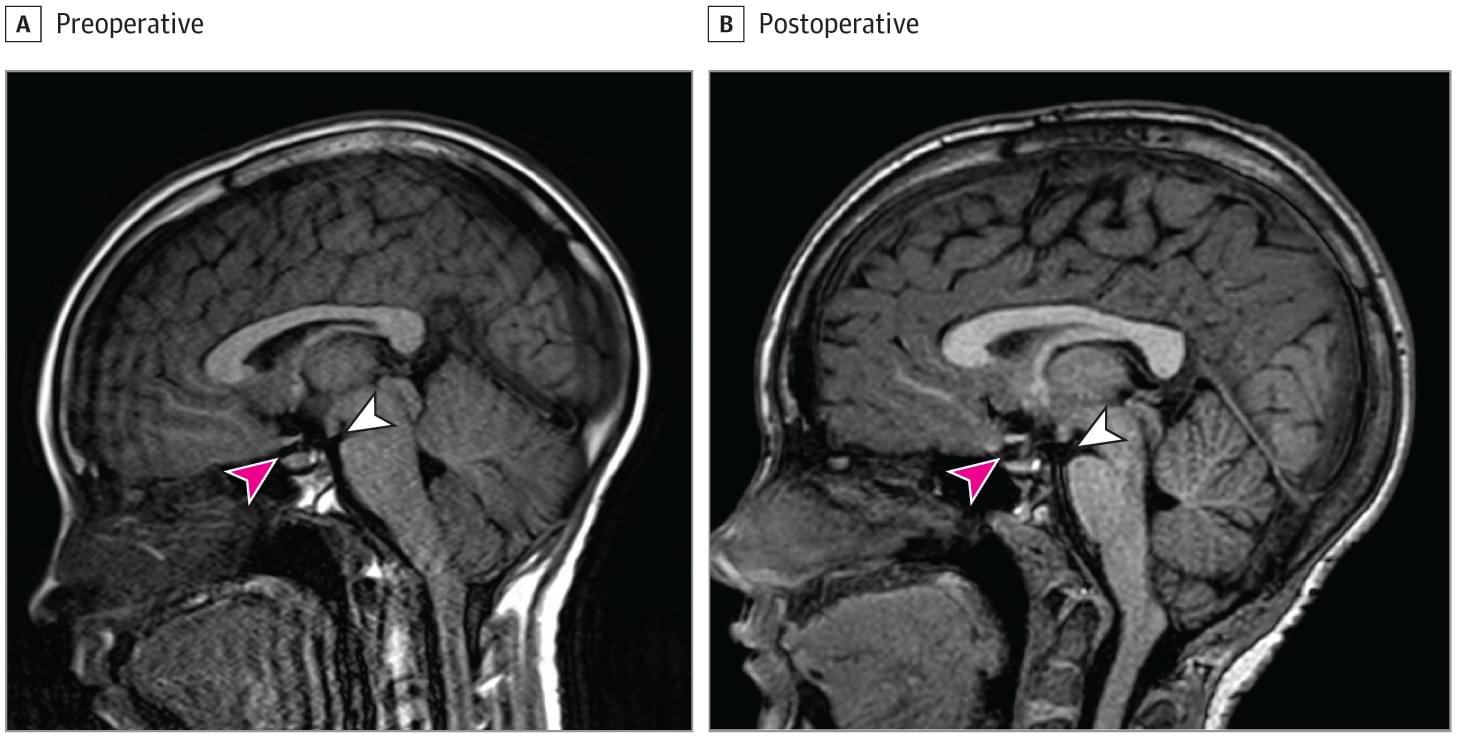
Spontaneous Intracranial Hypotension Due to Cerebrospinal Fluid Rhinorrhea
This case report describes an 8-year-old girl who presented with cerebrospinal fluid rhinorrhea, brain sagging, and orthostatic headaches.
T cells take up residence in the healthy brain via a gut-fat-brain axis
The brain is a unique place. It is shielded from much of the body by the blood-brain barrier, meaning it’s protected from pathogens and potentially dangerous substances that might be in our blood. And historically, many scientists believed that separation extended to the immune system as well: the brain has its own specialized immune cells called microglia, but immune cells present in the rest of the body were long thought to steer clear of the brain unless there was a disease or other problem requiring their presence.
Now, a team of scientists from Yale School of Medicine (YSM) has shown that immune cells known as T cells reside in the healthy brains of mice and humans, trafficked there from the gut and fat. This is the first time T cells have been shown to inhabit the brain under normal, non-diseased conditions.
The findings are published in Nature.