To explore how the brain deciphers the melody of speech, researchers worked with the rare group of patients who had electrodes implanted in their brains as part of epilepsy treatment. While these patients actively listened to an audiobook recording of “Alice in Wonderland,” scientists tracked activity in multiple brain regions in real time.
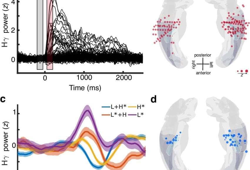
Using the intracerebral recordings from the electrodes deep in the patient’s brain, researchers noted the Heschl’s gyrus section processed subtle changes in voice pitch — not just as sound, but as meaningful linguistic units. The brain encoded pitch accents separately from the sounds that make up words.
The author says the research also revealed that the hidden layer of meaning carried by prosodic contours — the rise and fall of speech — is encoded much earlier in auditory processing than previously thought.
Similar research was conducted in non-human primates, but researchers found those brains lacked this abstraction, despite processing the same acoustic cues.
By unlocking the hidden layer of speech, the team discovered how the brain processes pitch accents, revealing profound implications for various fields.
“Our findings could transform speech rehabilitation, AI-powered voice assistants, and our understanding of what makes human communication unique,” the author said.