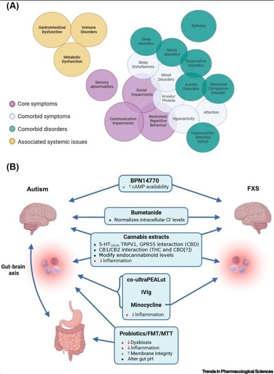
Toggle light / dark theme

Since 2014, the ALS Ice Bucket Challenge has inspired more than 17 million people to raise $115 million for The ALS Association, which has funded over 500 research projects with the money. Because of that boost, the first drug to treat ALS has been approved by the FDA, other new treatments are in testing, and scientists have been able to identify several genes that are connected to the disease.

While mutations in a gene called NEK1 have only been associated with around two percent of ALS cases, it is one of the primary genetic causes of ALS that have been revealed so far. Now investigators have learned more about how NEK1 mutations can lead to ALS, a disease in which the motor neurons that control movement degenerate and die, which causes paralysis and eventually, death. The work has been reported in Science Advances.

Researchers at the National Heart, Lung, and Blood Institute at NIH, Bethesda, have discovered a potential breakthrough for people with myalgic encephalomyelitis/chronic fatigue syndrome (ME/CFS), marked by extreme exhaustion, post-exertional malaise and cognitive issues.

In a paper, “WASF3 disrupts and may mediate exercise intolerance in /,” published in PNAS, the team details the influence of increased WASF3 proteins on the assembly of mitochondrial proteins, hampering energy production.

The study focused on a woman (S1) who experienced severe long-term fatigue. Measuring her muscles for phosphocreatine regeneration after exercise revealed a significant delay in mitochondrial ATP synthesis capacity. This discovery was followed up with a cell assay which found increased phospho-activation of an enzyme in a signaling pathway (MPAK).

I’d heard that fear of the dark is a protein, Scotophobin A, which can be isolated from the brains of rats. My Chemistry teacher told us that 1-hexanol smelled like cut grass. I watched her draw it once, on the whiteboard. A colorless liquid that, I imagined, smelled like memory, summer term, sports day, an army of ants cresting the summit of a picnic blanket, damp loam after rain.

I’d hoped that studying neuroscience would teach me all about things like that. I imagined watching sunlight refract through a conical flask, some clear liquid roiling inside. “Fear of abandonment is a sequence of seventeen peptides,” our lecturer might say, “isolated from the muscles of the heartbroken.”

“Look here,” he would say, pointing to another vial. “We can synthesize these things in a lab now. This one is awe.”

Scientists working in connectomics, a research field occupied with the reconstruction of neuronal networks in the brain, are aiming at completely mapping of the millions or billions of neurons found in mammalian brains. In spite of impressive advances in electron microscopy, the key bottleneck for connectomics is the amount of human labor required for the data analysis. Researchers at the Max Planck Institute for Brain Research in Frankfurt, Germany, have now developed reconstruction software that allows researchers to fly through the brain tissue at unprecedented speed. Together with the startup company scalable minds they created webKnossos, which turns researchers into brain pilots, gaining an about 10-fold speedup for data analysis in connectomics.

Billions of nerve cells are working in parallel inside our brains in order to achieve behaviours as impressive as hypothesizing, predicting, detecting, thinking. These neurons form a highly complex network, in which each nerve cell communicates with about one thousand others. Signals are sent along ultrathin cables, called axons, which are sent from each neuron to its about one thousand “followers.”

Only thanks to recent developments in , researchers can aim at mapping these networks in detail. The analysis of such image data, however, is still the key bottleneck in connectomics. Most interestingly, human annotators are still outperforming even the best computer-based analysis methods today. Scientists have to combine human and machine analysis to make sense of these huge image datasets obtained from the electron microscopes.

Seemingly countless self-help books and seminars tell you to tap into the right side of your brain to stimulate creativity. But forget the “right-brain” myth—a new study suggests it’s how well the two brain hemispheres communicate that sets highly creative people apart.

For the study, statisticians David Dunson of Duke University and Daniele Durante of the University of Padova analyzed the network of white matter connections among 68 separate brain regions in healthy college-age volunteers.

The brain’s white matter lies underneath the outer grey matter. It is composed of bundles of wires, or axons, which connect billions of neurons and carry electrical signals between them.

😗😁 Year 2022


Quantum processes are helpful to know about when we hear a gimcrack new theory that dismisses or explains away human consciousness. We know it can’t just be that simple.

You may also wish to read: Researchers: The brain’s claustrum acts as a router for thoughts Francis Crick thought the claustrum might be the “seat of consciousness,” an inherently materialist concept. The researchers think he was wrong. Of course, seeing the claustrum as a router is more consistent with the immaterial nature of consciousness than seeing it as a seat.

Glioblastoma is a fast-growing and aggressive brain tumor. As one of the most common malignant brain tumors, life expectancy after diagnosis is between 14 and 16 months. Roughly 1% of patients survive more than ten years with the longest patients living over 20 years. Symptoms include headaches, double vision, vomiting, loss of appetite, changes in mood and personality, inability to accurately think and learn, seizures, and difficulty speaking. Unfortunately, there is no cure, and treatment options include radiation and chemotherapy with limited efficacy. Glioblastoma is difficult to treat due to its location in the brain, its resistance to common treatment, the brains limited ability to heal itself, disrupted blood supply, blood vessel leakage, seizures, and neurotoxicity from treatments. Due to limited treatment and the life expectancy of this devastating disease, researchers at the SALK Institute in La Jolla, California have set out to find better ways to treat glioblastoma and prolong survival in patients.

Immune checkpoint inhibitors (ICIs) are a form of immunotherapy that block receptors on immune cells which activate them to kill tumor cells. The ICI using by the SALK group is known as anti-CTLA-4, which binds to the CTLA-4 protein on the T immune cells responsible for killing infected cells. This therapy was generated by Dr. James Allison at the MD Anderson Comprehensive Cancer Center in Houston, Texas. For his work, he was awarded the Nobel Prize in Physiology or Medicine in 2018. While this therapy proved effective in other cancers such as melanoma, it was unclear its effect in glioblastoma. The researchers at SALK recently published their findings on the effect of anti-CTLA-4 on glioblastoma.

The study published in Immunity by Dr. Susan Kaech and colleagues at SALK demonstrated prolonged survival of mice with glioblastoma after treatment with anti-CTLA-4. They also discovered that the treatment was largely dependent on CD4+ T cells, which aid in activating other cells, and not CD8+ T cells, which directly kill the tumor. More specifically, CD4+ T cells were found to infiltrate the brain and trigger other immune cells, like microglia to destroy cancerous cells. In Kaech’s work, the lab significantly shrunk the glioblastoma in mice and in some cases completely eradicated it.

Can you recognize someone you haven’t seen in years, but forget what you had for breakfast yesterday? Our brains constantly rearrange their circuitry to remember familiar faces or learn new skills, but the molecular basis of this process isn’t well understood. Today, scientists report that sulfate groups on complex sugar molecules called glycosaminoglycans (GAGs) affect “plasticity” in the brains of mice. Determining how GAGs function could help us understand how memory and learning work in humans, and provide ways to repair neural connectivity after injuries.

The researchers will present their results today at the fall meeting of the American Chemical Society (ACS).

The sugars that sweeten fruits, candies or cakes are actually just a few simple varieties of the many types of sugars that exist. When strung together, they can make a wide array of complex sugars. GAGs are formed by then attaching other chemical structures, including sulfate groups.

Exactly how, and how much, the unconscious processing of information influences our behavior has always been one of the most controversial questions in psychology. In the early 20th century, Sigmund Freud popularized the idea that our behaviors are driven by thoughts, feelings, and memories hidden deep within the unconscious mind — an idea that became hugely popular, but that was eventually dismissed as unscientific.

Modern neuroscience tells us that we are completely unaware of most brain activity, but that unconscious processing does indeed influence behavior; nevertheless, certain effects, such as unconscious semantic “priming,” have been called into question, leading some to conclude that the extent of unconscious processing is limited.

A recent brain scanning study now shows that unconsciously processed visual information is distributed to a wider network of brain regions involved in higher-order cognitive tasks. The results contribute to the debate over the extent to which unconscious information processing influence the brain and behavior and led the authors of the study to revise one of the leading theories of consciousness.