Flatworms can rebuild themselves from just a small fragment, and now scientists know why. Their stem cells ignore nearby instructions and respond to long-distance signals from other tissues. This discovery turns old stem cell theories upside down and could lead to new ways to repair or regrow human tissue. It also reveals a hidden complexity in one of nature’s simplest creatures.
Category: life extension – Page 34


Drug combo extends life of frail male mice by ‘remarkable’ 73%, scientists find
“Compared to other established lifespan-extending interventions, Oxytocin+A5i demonstrates unique outcomes, such as significantly (over 70 per cent) increased life expectancy from the start of this therapy in old and frail male mice, and a robust decrease in mortality risk,” they wrote.
The latest study also found that the Oxytocin+A5i treatment reduced chaotic levels of some circulating blood proteins, which are key markers of ageing, bringing their levels back to a more youthful state.
However, after four months of continuous treatment, only male mice showed sustained improvement in these protein levels.

Scientists Can Now “See” Aging Through Your Eyes
The small blood vessels in the eye could reveal important clues about a person’s risk of heart disease and the rate at which they are biologically aging, according to scientists from McMaster University and the Population Health Research Institute (PHRI) – a joint institute of Hamilton Health Sciences and McMaster.
Published in the journal Science Advances, the research suggests that retinal scans may eventually become a simple, non-invasive way to assess the body’s vascular health and aging process. This approach could pave the way for earlier detection of health issues and more effective preventive care.
“By connecting retinal scans, genetics, and blood biomarkers, we have uncovered molecular pathways that help explain how aging affects the vascular system,” says Marie Pigeyre, senior author of the study and associate professor with McMaster’s Department of Medicine.


Isotropic MOF coating reduces side reactions to boost stability of solid-state Na batteries
In recent years, energy engineers have been trying to design new reliable batteries that can store more energy and allow electronics to operate for longer periods of time before they need to be charged. Some of the most promising among these newly developed batteries are solid-state batteries, which contain solid electrolytes instead of liquid ones.
Compared to batteries with liquid electrolytes that are widely used today, solid-state batteries could exhibit higher energy densities (i.e., could store more energy) and longer lifetimes. However, many of these batteries have been found to be unstable, due to unwanted chemical reactions that occur between their high-voltage cathodes (i.e., positive electrodes) and solid electrolytes, which can speed up the degradation of the batteries’ performance over time.
These undesirable side reactions are particularly common in sodium-ion (Na+) solid-state batteries, which use Na+ ions to store and release electrical energy. This is because while Na is more abundant and cheaper than lithium, Na-ion batteries are inherently more chemically reactive than Li-ion batteries.

Microglial gene mutation inked to increased Alzheimer’s risk
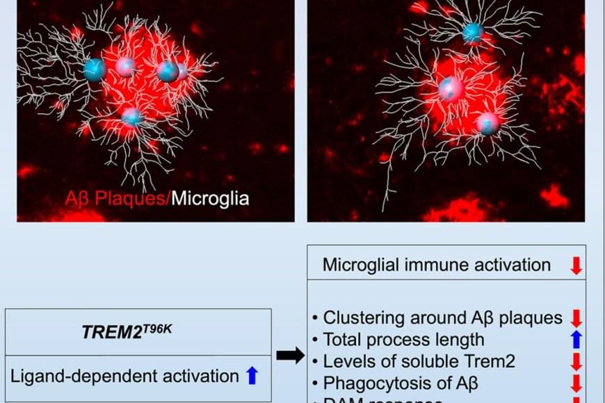
The team wanted to understand how immune cells of the brain, called microglia, contribute to Alzheimer’s disease (AD) pathology. It’s known that subtle changes, or mutations, in genes expressed in microglia are associated with an increased risk for developing late-onset AD.
The study focused on one such mutation in the microglial gene TREM2, an essential switch that activates microglia to clean up toxic amyloid plaques (abnormal protein deposits) that build up between nerve cells in the brain. This mutation, called T96K, is a “gain-of-function” mutation in TREM2, meaning it increases TREM2 activation and allows the gene to remain super active.
They explored how this mutation impacts microglial function to increase risk for AD. The authors generated a mutant mouse model carrying the mutation, which was bred with a mouse model of AD to have brain changes consistent with AD. They found that in female AD mice exclusively, the mutation strongly reduced the capability of microglia to respond to toxic amyloid plaques, making these cells less protective against brain aging.

Scientists find ways to boost memory in aging brains
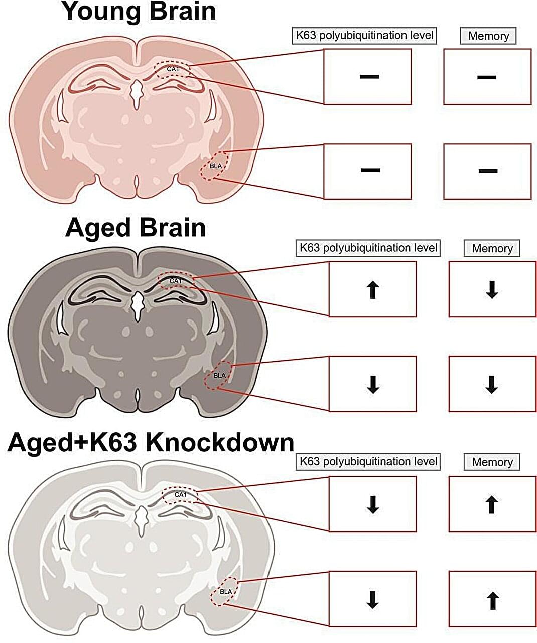
Memory loss may not simply be a symptom of getting older. New research from Virginia Tech shows that it’s tied to specific molecular changes in the brain and that adjusting those processes can improve memory.
In two complementary studies, Timothy Jarome, associate professor in the College of Agriculture and Life Sciences’ School of Animal Sciences, and his graduate students used gene-editing tools to target those age-related changes to improve memory performance in older subjects. The work was conducted on rats, a standard model for studying how memory changes with age.
“Memory loss affects more than a third of people over 70, and it’s a major risk factor for Alzheimer’s disease,” said Jarome, who also holds an appointment in the School of Neuroscience. “This work shows that memory decline is linked to specific molecular changes that can be targeted and studied. If we can understand what’s driving it at the molecular level, we can start to understand what goes wrong in dementia and eventually use that knowledge to guide new approaches to treatment.”

The Rise of Mechanobiology for Advanced Cell Engineering and Manufacturing
The rise of cell-based therapies, regenerative medicine, and synthetic biology, has created an urgent need for efficient cell engineering, which involves the manipulation of cells for specific purposes. This demand is driven by breakthroughs in cell manufacturing, from fundamental research to clinical therapies. These innovations have come with a deeper understanding of developmental biology, continued optimization of mechanobiological processes and platforms, and the deployment of advanced biotechnological approaches. Induced pluripotent stem cells and immunotherapies like chimeric antigen receptor T cells enable personalized, scalable treatments for regenerative medicine and diseases beyond oncology. But continued development of cell manufacturing and its concomitant clinical advances is hindered by limitations in the production, efficiency, safety, regulation, cost-effectiveness, and scalability of current manufacturing routes. Here, recent developments are examined in cell engineering, with particular emphasis on mechanical aspects, including biomaterial design, the use of mechanical confinement, and the application of micro-and nanotechnologies in the efficient production of enhanced cells. Emerging approaches are described along each of these avenues based on state-of-the-art fundamental mechanobiology. It is called on the field to consider mechanical cues, often overlooked in cell manufacturing, as key tools to augment or, at times, even to replace the use of traditional soluble factors.
Current manufacturing workflows for CAR-based immunotherapies, particularly CAR T, and the emerging CAR NK and CAR macrophage platforms, generally involve four key stages: (i) isolation of primary immune cells or their precursors, (ii) cell activation or differentiation, (iii) genetic modification with CAR constructs, most often via viral vectors or electroporation (EP), and (iv) expansion or preparation for reinfusion. Among these, transfection remains the most critical and technically challenging step, directly influencing the functionality, safety, and scalability of the final product.
In clinical-scale production, EP remains the most widely used non-viral method for gene delivery into immune cells, yet it is increasingly recognized as suboptimal, particularly when delivering large or complex CAR constructs. It suffers from inefficient nuclear delivery, high cell toxicity, and poor functional yields of viable, potent CAR-expressing cells.[ 113 ] These limitations are further exacerbated in more fragile or less permissive cell types, such as NK cells and macrophages, which show lower transfection efficiencies and greater sensitivity to electroporation-induced stress.[ 114 ] Viral vectors, while still dominant in clinical manufacturing, present their own challenges: they are constrained by limited cargo capacity, are costly to produce at scale, and raise regulatory and safety concerns, especially when applied to emerging CAR-NK and CAR macrophage therapies that require flexible, transient, or multiplexed genetic programs.[ 115 ]
In contrast to immune-cell engineering, stem cell-based approaches present a different set of challenges and engineering requirements. While immune cells are genetically modified to enhance cytotoxicity[ 116 ] and specificity or to mitigate excessive T-cell activation,[ 117 ] stem cells must be engineered to control self-renewal, lineage commitment, and functional integration, often requiring precise, non-integrative delivery of genetic or epigenetic modulators (e.g., mRNA, episomal vectors) to maintain cellular identity and safety.[ 118 ] Stem cells hold exceptional therapeutic promise due to their capacity for self-renewal and differentiation into specialized cell types, supporting applications in personalized disease modeling, tissue repair, and organ regeneration.[ 119 ] However, engineering stem cells in a safe, efficient, and clinically relevant manner remains a major challenge. Conventional delivery methods, such as viral vectors and EP, can compromise genomic integrity,[ 120 ] reduce viability,[ 118 ] and induce epigenetic instability,[ 121 ] limiting their translational potential.