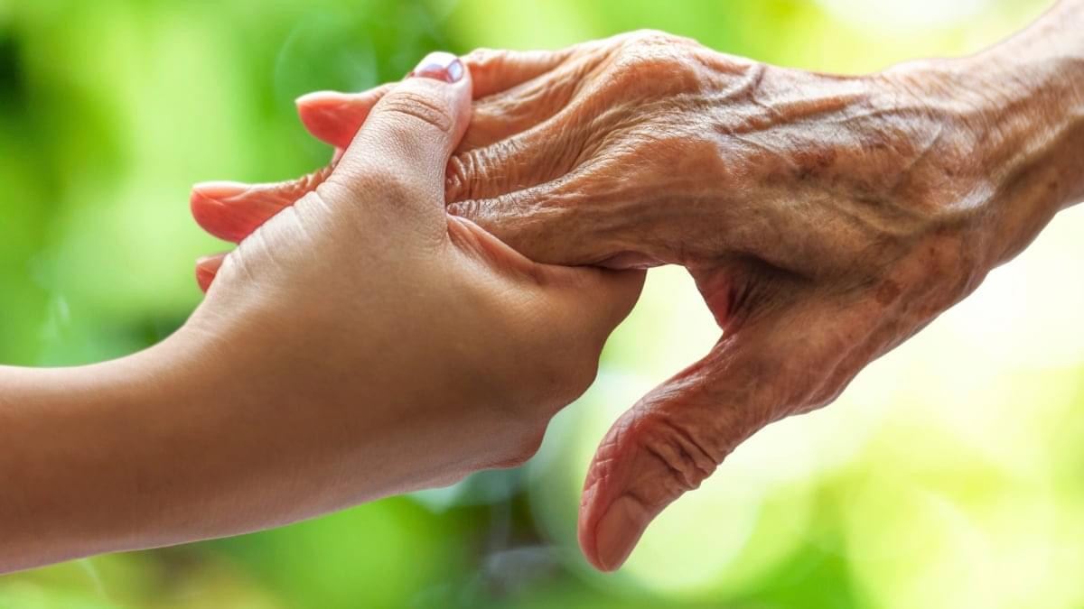
In this groundbreaking conversation, Professor of Genetics and longevity scientist, Dr. David Sinclair, A.O., Ph.D., joins Sarah Grynberg to unpack the future of human aging, the science of longevity, and how we live today impacts how we age tomorrow.
From reversing blindness in mice to exploring treatments that could one day delay menopause and extend healthy human life, this episode will completely change the way you think about your body, your health, and your future.
But beyond the science, this is also a deeply human conversation about purpose, suffering, love, family, and what it truly means to live a great life.
In this episode, you will learn:
Why aging may actually be reversible.
The daily habits accelerating aging in your body right now.
How stress, loneliness, and cortisol could impact longevity.
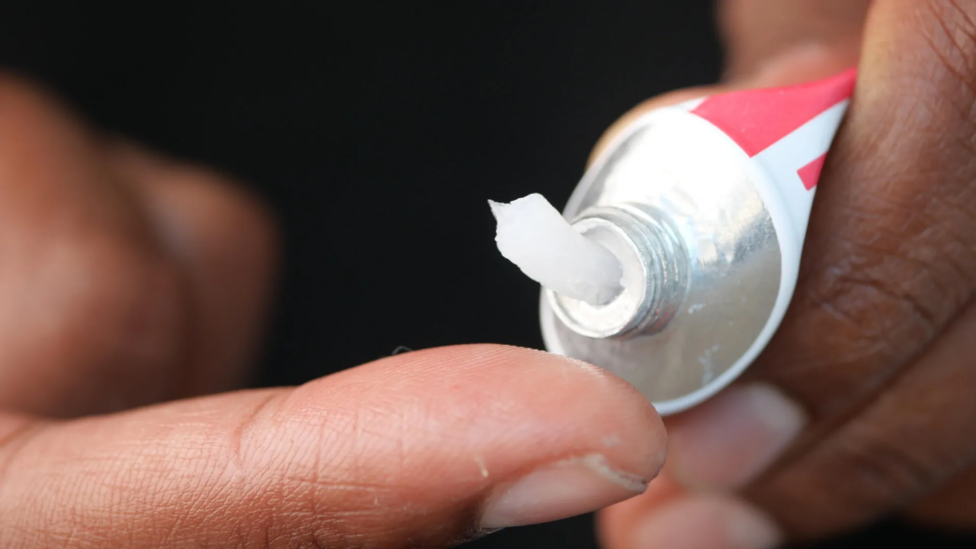
The real science behind supplements like NMN, resveratrol, and NAD boosters.
Why exercise, sleep, and relationships matter more than you think.
What Dr. Sinclair believes is coming in the next 10 years of medicine.
How scientists are working to reverse female infertility and delay menopause.
The surprising reason your “biological age” may be younger or older than your real age.
Why suffering through disease and decline should not be considered “normal aging”
The philosophy and mindset Dr. Sinclair lives by every day.
00:00 — Introduction.
01:18 — Why David Sinclair Became Obsessed With Aging.
06:20 — The Childhood Conversation That Changed His Life.
10:18 — The Groundbreaking Discovery That Could Reverse Aging.
12:47 — Reversing Blindness In Mice.
13:33 — Human Trials Are About To Begin.
16:11 — What Accelerates Aging Faster Than Anything Else.
20:08 — Why Relationships & Loneliness Impact Longevity.
24:14 — The Truth About Sun Exposure & Aging.
28:59 — Alzheimer’s, Cancer & Diseases Of Aging.
35:28 — Will Humans Live Longer In The Next Decade?
38:34 — The Supplements David Sinclair Personally Takes.
46:50 — Menopause, Fertility & Reversing Ovarian Aging.
50:20 — What Humans Will Eventually Die From.
51:18 — The Difference Between His Mother & Father’s Aging.
55:37 — Skin Rejuvenation, Hair Growth & Looking Younger.
58:16 — Why He Became A “Struggling Vegan”
01:00:08 — David Sinclair’s Workout & Exercise Routine.
01:03:28 — The Lifespan Community & Podcast.
01:06:02 — The Best Advice He’s Ever Received.
01:08:09 — What A Life Of Greatness Means To David Sinclair.
This episode is a powerful reminder that longevity is not just about living longer… it’s about living better.

