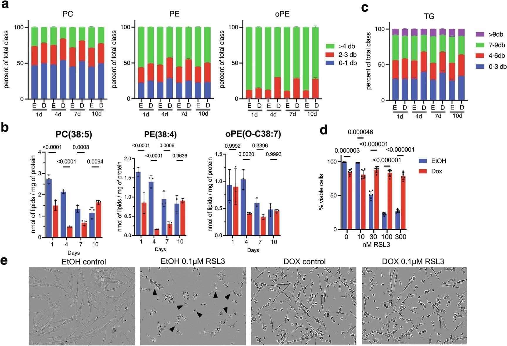
In response to stress or damage, cells undergo senescence and stop dividing. However, if senescent cells accumulate in tissues over the long term, chronic inflammation occurs and the risk of cancer increases. Researchers at the German Cancer Research Center (DKFZ) have now discovered a previously unknown mechanism by which senescent cells protect themselves from oxidative stress and a specific form of cell death known as ferroptosis.
In the long term, these findings could provide new avenues for cancer therapies and the treatment of age-related diseases. The research is published in the journal Cell Death & Differentiation.
Senescence occurs when cells respond to stress or harmful changes and permanently cease their growth. This process is considered a protective mechanism against cancer. For example, cells that carry an oncogene permanently activated by mutations are effectively “frozen” before they can proliferate uncontrollably—a biological emergency program. However, problems arise when senescent cells accumulate in tissue, where they promote chronic inflammation and thus facilitate tumor development. Scientists are therefore searching for ways to eliminate senescent cells before they can cause harm.
A cellular explanation for how tau aggregates into fibrils in Alzheimer’s disease has been elusive. This paper identifies the failure of ‘neuroproteasomes’ as sufficient to convert tau into paired helical filaments, a process regulated by ApoE and aging.
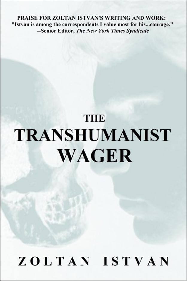
Thirteen years ago, I sat down with a writer who had just published his first novel.
It was Zoltan Istvan’s very first media interview as a book author.
The book was The Transhumanist Wager. The question behind it was simple and almost unbearable: what would you do, and what would you give up, to live forever?
I loved half of it. I argued with the other half. That tension is exactly why I think it still matters.
Zoltan built his story out of Plato and Nietzsche, out of Thomas More’s Utopia and Zen Buddhism, then wrapped it all in an Atlas Shrugged plot of lone heroes and evil states. The philosophy is sophisticated. The framing is stark. The contradictions are not a flaw. They are the point.
One line from our conversation has stayed with me for more than a decade:
New LDL Drug Could Cure Heart Disease. Eli Lilly published Phase 1 data in the New England Journal of Medicine showing that a single IV infusion of a gene editing therapy called VERVE-102 lowered LDL cholesterol permanently. Well… the effect held for at least 18 months. Longevity Twitter immediately called it the cure for heart disease. The science is real. The hype is getting ahead of what the paper actually says. This episode walks through the Phase 1 Heart-2 trial — the data, the base-editing mechanism (which is NOT CRISPR), the one safety event nobody’s talking about, and how Eli Lilly’s CEO is publicly thinking about pricing a one-and-done cure.
HUME BODY POD DISCOUNT UP TO 50% OFF: Code: LSN20 https://humehealth.com/pages/hume-bod… Latte: https://longevitylatte.shop TIMESTAMPS 0:00 — Cold Open 1:41 — Sponsor: Hume Body Pod 2:53 — Intro: The Cholesterol Paradox 4:48 — How Statins Work 5:36 — PCSK9 Targeting 7:38 — How VERVE-102 Works 9:16 — The Results 12:16 — Other Drugs and Pricing 15:12 — Natural Alternatives 17:42 — Other One and Done Drugs 18:40 — Longevity Latte SOURCES & LINKS NEJM paper (Vafai, Täubel, Patel, Kathiresan et al.): https://www.nejm.org/doi/full/10.1056… Eli Lilly press release on Phase 1 Heart-2 data: https://www.prnewswire.com/news-relea… ClinicalTrials.gov Heart-2 trial entry (NCT06164730): https://clinicaltrials.gov/study/NCT0… Verve Therapeutics FDA Fast Track designation announcement: https://vervetx.gcs-web.com/news-rele… Cohen and Hobbs 2006 NEJM paper (the foundational PCSK9 loss-of-function discovery): https://www.nejm.org/doi/abs/10.1056/.… Dave Ricks (Eli Lilly CEO) on Cheeky Pint with Patrick and John Collison: • Dave Ricks, CEO of Eli Lilly, on GLP-1s an… FOURIER trial (evolocumab cardiovascular outcomes): https://www.nejm.org/doi/full/10.1056… ODYSSEY OUTCOMES trial (alirocumab cardiovascular outcomes): https://www.nejm.org/doi/full/10.1056… PCSK9 LoF and diabetes (Mendelian randomization, Lancet Diabetes & Endocrinology): https://www.thelancet.com/journals/la… PCSK9 inhibition and diabetes risk review: https://pmc.ncbi.nlm.nih.gov/articles… StatPearls overview of PCSK9 inhibitors: https://www.ncbi.nlm.nih.gov/books/NB… Sardinia cholesterol paradox study: https://pmc.ncbi.nlm.nih.gov/articles… Statin pleiotropic effects review (mevalonate pathway): https://www.ncbi.nlm.nih.gov/pmc/arti… Berberine as a nature-made PCSK9 inhibitor review: https://www.ncbi.nlm.nih.gov/pmc/arti… Berberine for dyslipidaemias meta-analysis: https://pubmed.ncbi.nlm.nih.gov/30466… Pomegranate juice, carotid IMT, and LDL oxidation (Aviram 3-year study): https://www.clinicalnutritionjournal… LATEST EXCLUSIVE INTERVIEW: [Ariel Garten / Muse headset interview YouTube URL] ABOUT LONGEVITY SCIENCE NEWS Longevity Science News covers the latest breakthroughs in anti-aging research, regenerative medicine, longevity biotech, and the science of extending human healthspan and lifespan. Hosted by Emmett Short. Disclaimer: This video is for educational and informational purposes only and does not constitute medical advice. Consult a qualified clinician before making health or treatment decisions. EXCLUSIVE INTERVIEWS & BONUS CONTENT Patreon: https://patreon.com/u29506604?utm_med… YT Membership: / @longevitysciencenews PRODUCTION CREDITS Executive Producer – Keith Comito Host, Producer, Writer – Emmett Short.
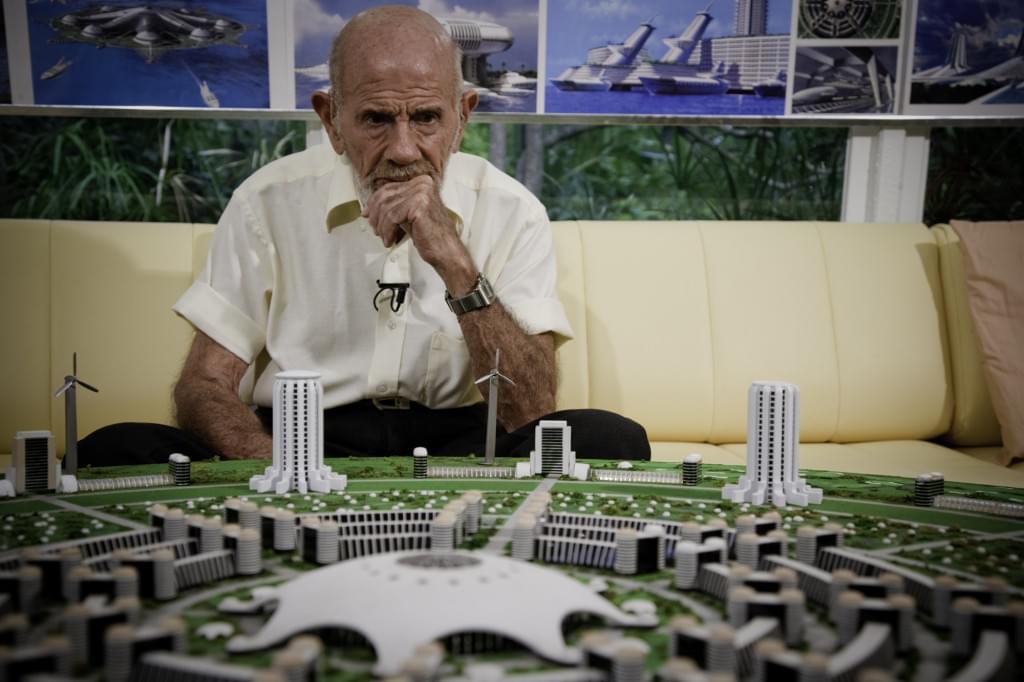
I really liked Jacque Fresco. Not as a thinker I was supposed to admire, but as a person: the humor, the humility, the scientific curiosity still burning at 97.
That made the disagreements harder, not easier.
Fresco spent almost a century arguing one idea. We apply the methods of #science to engineering, to medicine, to flight. Then we run our economies and our politics on opinion, tradition, and the preferences of the financial elite.
He thought we had it exactly inverted. Rigor for the machines, guesswork for the humans.
“Technology was never the hard part. The harder question is what kind of society we want it to serve.”
Hackers are ruthless. They can take control of your computer, delete files and disappear without a trace. However, FIU cybersecurity researcher Weidong Zhu has discovered a way to transform a computer’s storage chip into an additional tool for cyber defense. Working with collaborators at the University of Florida, Zhu created a system that makes data on these chips last longer—extending the lifespan of your files in the critical window after your computer is compromised. The work is published in the journal Proceedings of the 2025 ACM SIGSAC Conference on Computer and Communications Security.
“Our system extends recoverable data history up to 126 days,” said Zhu, an assistant professor at FIU’s Knight Foundation School of Computing & Information Sciences whose work is part of the Center for Integrated Security, Privacy, and Trustworthy AI (CIERTA). “Even if your computer is infected, your data can survive on your drive.”
Storage chips, known as solid-state drives (SSDs), have intrigued cybersecurity researchers for years. As hardware—not software—they offer unique safety benefits during an attack.
Aging is no longer viewed as an untouchable part of life. According to Eric Verdin, scientists are beginning to treat aging itself as a biological process that can be slowed and potentially reversed.
In this episode, Eric explains why longevity research is entering a new era. He discusses how AI, women’s health, metabolic therapies, and partial reprogramming are reshaping medicine. He highlights GLP-1 drugs as one of the most promising tools today and explains how resetting cells to a younger state may one day restore function in aging tissues.
He also shares the most effective strategies available right now: exercise, sleep, nutrition, mental stimulation, and social connection. While supplements like Creatine may help, Eric stresses that lifestyle remains the foundation of long-term health.
Eric Verdin is a physician-scientist and CEO of the Buck Institute for Research on Aging, where he leads research focused on extending human healthspan.
From time to time, Harvard Health Publishing issues Special Health Reports – consumer-facing, doctor-reviewed guides translating medical research for general readers. Previous reports included topics such as Alzheimer’s and heart disease. This new one, presented to the public earlier this week, is dedicated to healthy longevity. While this report, aimed mostly at curious laypeople and priced at $29, might not reveal a trove of new information to a longevity-savvy reader, it is an unmistakable sign that longevity science and the very idea of extending lifespan and healthspan are finally entering the mainstream.
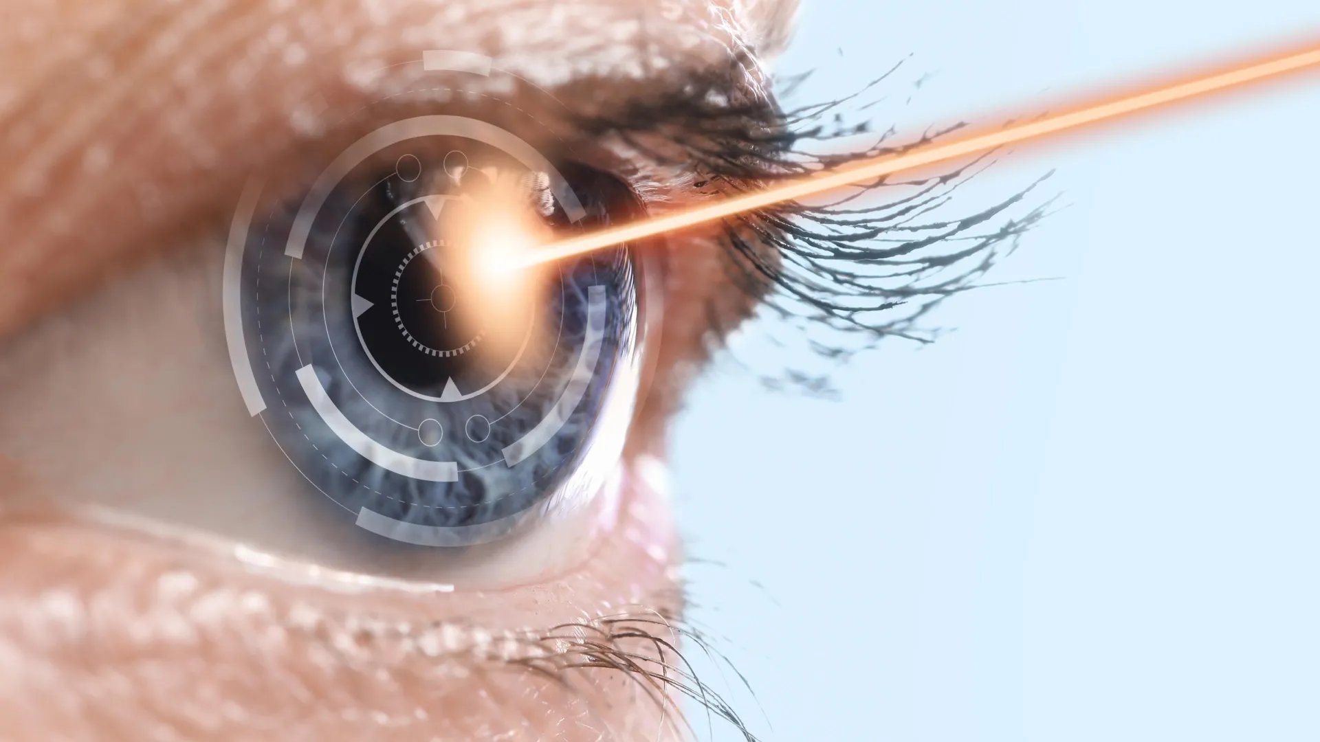
A new experimental treatment could finally offer hope for millions of people with dry age-related macular degeneration — one of the leading causes of blindness in older adults. Researchers at Aalto University discovered a way to gently heat tissue at the back of the eye using near-infrared light, triggering the cells’ natural “cleanup and repair” systems before major damage occurs.


