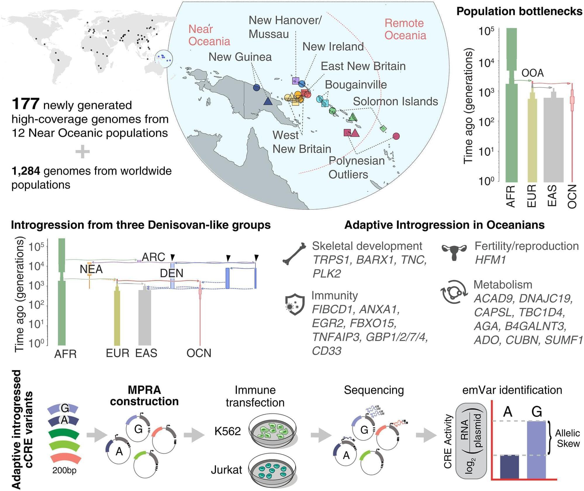
Toggle light / dark theme

Brain motion is driven by mechanical coupling with the abdomen

UNIVERSITY PARK, Pa. — The brain is more mechanically connected to the body than previously appreciated, scientists reported today (April 27) in Nature Neuroscience. Through a study using mice and simulations, the team found a potential biological mechanism underlying why exercise is thought to benefit brain health: abdominal contractions compress blood vessels connected to the spinal cord and the brain, enabling the organ to gently move within the skull. This swaying facilitates the surrounding cerebrospinal fluid to flow over the brain, potentially washing away neural waste that could cause problems for brain function.

According to Patrick Drew, professor of engineering science and mechanics, of neurosurgery, of biology and of biomedical engineering at Penn State, the work builds on previous studies detailing how sleep and neuron loss can influence how and when cerebrospinal fluid flushes through the brain.

“Our research explains how just moving around might serve as an important physiological mechanism promoting brain health,” said Drew, corresponding author on the paper. “In this study, we found that when the abdominal muscles contract, they push blood from the abdomen into the spinal cord, just like in a hydraulic system, applying pressure to the brain and making it move. Simulations show that this gentle brain movement will drive fluid flow in and around the brain. It is thought the movement of fluid in the brain is important for removing waste and preventing neurodegenerative disorders. Our research shows that a little bit of motion is good, and it could be another reason why exercise is good for our brain health.”

Drew, who also holds the title of associate director of the Huck Institutes of the Life Sciences, explained how in a hydraulic system, a pump creates pressure that drives fluid flow. In this case, the pump is the abdominal contraction — which can be as light as the tensing prior to sitting up or taking a step. The contraction puts pressure on the vertebral venous plexus, a network of veins that connect the abdominal cavity to the spinal cavity, causing the brain to move.

Abstract: Nature Neuroscience Brain motion is driven by mechanical coupling with the abdomen.

Read More


Human DNA-PKcs promotes broken DNA-end structure independence during NHEJ

Whether DNA-PKcs is necessary for non-homologous end joining has been biochemically obscure. Through optimization of reaction conditions, Fujii and Modesti show that DNA-PKcs plays a constructive role, which leads to indistinguishable repair efficiencies between cohesive-end and blunt-end DNA substrates.

Abstract: Metabolic syndrome and excessive alcohol consumption lead to liver injury and fibrosis, characterized by increased collagen deposition from hepatic stellate cells

https://doi.org/10.1172/JCI197923 Here, David A. Brenner & team discover the RNA-binding protein LARP6 as a master coordinator of hepatic stellate cell activation and fibrosis, using human tissue and liver spheroid models of MASH and MetALD.

The image shows collagen labeling (red) in human liver spheroids MASH model with LARP6-targeting ASO (DAPI, blue). Collagen labeling is decreased compared with MASH control.


11 Center for Epigenomics, UCSD, La Jolla, California, USA.

12 Sanford Burnham Prebys Medical Discovery Institute, La Jolla, California, USA.

13 Department of Biomedical Sciences, College of Medicine, Florida State University, Tallahassee, Florida, USA.

Experiments advance efforts to restore vision with transplanted neurons

Researchers at Johns Hopkins Medicine say they have successfully demonstrated that disrupting an eye structure long suspected of blocking the growth and survival of transplanted nerve cells may help restore vision in people with optic nerve damage.

A report on the experiments with animals, stem cells and donated eye tissue was published in Science Translational Medicine. It suggests that altering or removing a thin layer of tissue called the internal limiting membrane, which separates the light-sensing retinal tissue at the back of the eye from the gel-like vitreous fluid that fills the eye, could help transplanted retinal ganglion cells (RGCs) survive and grow in people with blinding optic nerve damage.

Such damage, also known as optic neuropathy, occurs when retinal ganglion cells die of disease, inflammation or injury and stop carrying electrical signals to the brain. Common causes of damage include glaucoma, optic nerve inflammation (optic neuritis) and ischemic optic neuropathy (sudden loss of blood flow to the optic nerve).

Neuromodulation for gait disorders

Gait impairments such as freezing, weakness and imbalance remain resistant to standard therapies across neurological disorders. This Review examines advances in neuromodulation, from refining deep brain stimulation to integrating spinal and distributed strategies. It discusses adaptive neurotechnologies, mechanistic insights and a framework for tailoring spatiotemporally precise interventions to restore gait control.

New sensor sniffs out pneumonia on a patient’s breath

Diagnosing some diseases could be as easy as breathing into a tube. MIT engineers have developed a test to detect disease-related compounds in a patient’s breath. The new test could provide a faster way to diagnose pneumonia and other lung conditions. Rather than sit for a chest X-ray or wait hours for a lab result, a patient may one day take a breath test and get a diagnosis within minutes.

The new breath test is a portable, chip-scale sensor that traps and detects synthetic compounds, or “biomarkers,” of disease, which are initially attached to inhalable nanoparticles. The biomarkers serve as tiny tags that can only be unlocked and detached from the nanoparticle by a very particular key, such as a disease-related enzyme.

The idea is that a person would first breathe in the nanoparticles, similar to inhaling asthma medicine. If the person is healthy, the nanoparticles would eventually circulate out of the body intact. If a disease such as pneumonia is present, however, enzymes produced as a result of the infection would snip off the nanoparticles’ biomarkers. These untethered biomarkers would be exhaled and measured, confirming the presence of the disease.

/* */