Research by the Barcelona Institute for Biomedical Research (IIBB), part of the Spanish National Research Council (CSIC), and the Institut de Recerca Sant Pau (IR Sant Pau) provides some of the first evidence that psychological therapies act as biological stimuli that induce molecular responses measurable through blood biomarkers.
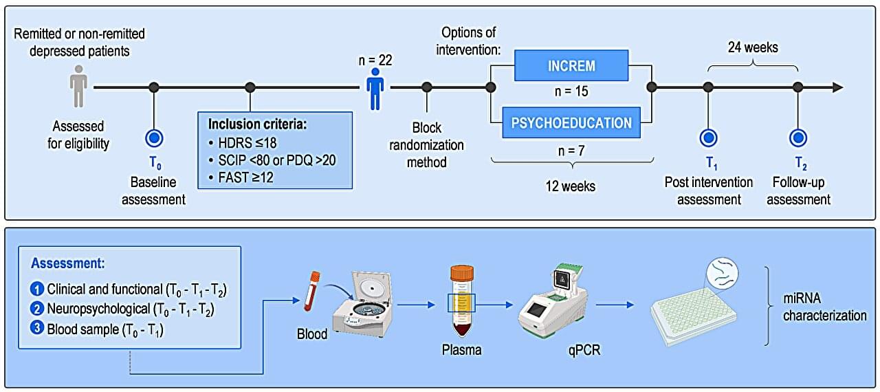
The preliminary study, involving 22 patients with major depressive disorder at Hospital de Sant Pau, reveals that psychotherapy sessions trigger changes in microRNAs—molecules that regulate gene expression in cells—associated with significant improvements in the participants’ cognitive status. The results, published in Scientific Reports, represent an advance toward monitoring patients’ responses to pharmacological treatments and nonpharmacological therapeutic interventions.
The study, led by Dr. Maria J. Portella (IR Sant Pau) and Dr. Analia Bortolozzi (IIBB-CSIC), with Lluís Miquel-Rio (IIBB-CSIC) and Dr. Muriel Vicent-Gil (Hospital de Sant Pau) as first authors, focused on major depressive disorder (MDD). This condition is characterized not only by its effects on mood but also by a broad spectrum of cognitive impairments, including difficulties with attention, memory, processing speed and executive function. These symptoms frequently persist despite treatment and severely affect patients’ quality of life.
