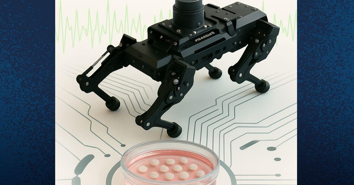
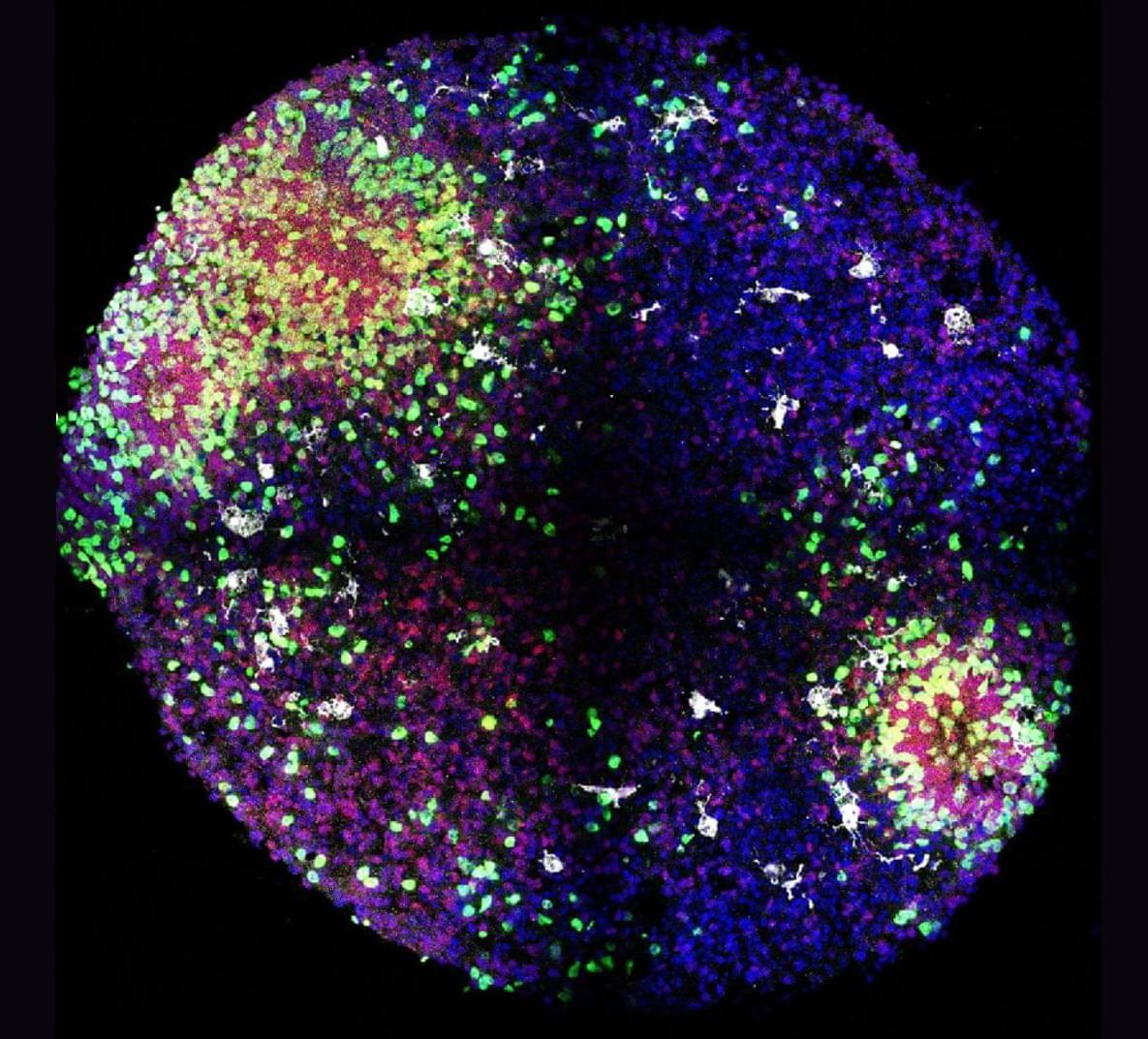
Toggle light / dark theme

Scientists Tweaked LSD’s Molecular Structure and Created a Wild New Brain Drug

A team of researchers at the University of California, Davis, made small tweaks to the molecular structure of lysergic acid diethylamide (LSD) to see if it could be turned into an effective brain-healing treatment for patients that suffer from conditions like schizophrenia — without risking a potentially disastrous acid trip.

As detailed in a new paper published in the journal Proceedings of the National Academy of Sciences last month, the researchers created a new compound called JRT by shifting the position of just two atoms of the psychedelic’s molecular structure.

With the two atoms flipped, the new drug could still stimulate brain cell growth and repair damaged neural connections, while simultaneously minimizing psychedelic effects, in mice.

Common analgesic gas aids in opening of blood-brain barrier

Nitrous oxide, a commonly used analgesic gas, temporarily improved the opening of the blood-brain barrier (BBB) to allow gene therapy delivery in mouse models using focused ultrasound (FUS), UT Southwestern Medical Center researchers report in a new study. Their findings, published in Gene Therapy, could eventually lead to new ways to treat a variety of brain diseases and disorders.

“The approach we explored in this study has the potential to advance care for diseases of the brain that can be treated by targeted therapeutic delivery,” said study leader Bhavya R. Shah, M.D., Associate Professor of Radiology, Neurological Surgery, and in the Advanced Imaging Research Center at UT Southwestern. He’s also an Investigator in the Peter O’Donnell Jr. Brain Institute and a member of the Center for Alzheimer’s and Neurodegenerative Diseases. Deepshikha Bhardwaj, Ph.D., Senior Research Associate at UTSW, was the study’s first author.

The BBB is a highly selective border of semipermeable cells that line tiny blood vessels supplying blood to the brain. It is thought to have developed during evolution to protect the brain from toxins and infections in the blood. However, the BBB also impedes the delivery of drugs that could be used to treat neurologic or neuropsychiatric conditions, such as Alzheimer’s disease, multiple sclerosis, or brain tumors. Consequently, researchers have worked for decades to develop solutions that can temporarily open the BBB to allow treatments to enter.

«Matrix: Start» — researchers powered 350 LEDs from the human body

Researchers at the University of Alabama in the United States have used duct tape from a store to create a triboelectric nanogenerator capable of collect electricity from the human body and the environment.

Their development is capable of powering small devices such as biosensors by converting mechanical energy from friction and movement into electricity. The generator is made of metallized films polyethylene terephthalate, which act as electrodes, and layers of adhesive tape.

The developers emphasize that power generation occurs through interaction polypropylene and of the acrylic adhesive layer when they are pressed and released. At the same time, due to the weak intermolecular interaction (Van der Waals forces) on the borders of the atomic-sized gaps.

Flexible imager that’s thinner than an eyelash can capture brain activity

Researchers have developed an extremely thin, flexible imager that could be useful for noninvasively acquiring images from inside the body. The new technology could one day enable early and precise disease detection, providing critical insights to guide timely and effective treatment.

“As opposed to existing prohibitively large endoscopes made of cameras and or bulky fiber optic bundles, our microimager is very compact,” said research team leader Maysam Chamanzar from Carnegie Mellon University. “Much thinner than a typical eyelash, our device is ideal for reaching deep regions of the body without causing significant damage to the tissue.”

In the journal Biomedical Optics Express, the researchers showed that the microimager, which is only 7 microns thick—a tenth of an eyelash diameter—and about 10 mm long, can be used in a for structural and functional imaging of brain activity. The width of the thin film imager can be customized based on the desired field of view and resolution.

Human-specific DNA enhancer linked to brain development and neuron proliferation

Duke University Medical Center-led research has identified a human-specific DNA enhancer that regulates neural progenitor proliferation and cortical size. Small genetic changes in HARE5 amplify a key developmental pathway, resulting in increased cortical size and neuron number in experimental models. Findings have implications for understanding the genetic mechanisms underlying neurodevelopmental disorders.

Humans possess a significantly larger and more complex cerebral cortex compared to other species, contributing to advanced cognitive functions. Comparative genomics research has identified Human Accelerated Regions (HARs), segments of non-coding DNA with human-specific genetic changes. Many HARs are located near genes associated with and neural differentiation.

Because thousands of HARs have been identified and linked to brain-related genes, the next critical step is to investigate how these actively shape human brain features.

Computational strategy reveals potential new targets for Alzheimer’s drugs

The study revealed genes and cellular pathways that haven’t been linked to Alzheimer’s before, including one involved in DNA repair. Identifying new drug targets is critical because many of the Alzheimer’s drugs that have been developed to this point haven’t been as successful as hoped.

Working with researchers at Harvard Medical School, the team used data from humans and to identify cellular pathways linked to neurodegeneration. This allowed them to identify additional pathways that may be contributing to the development of Alzheimer’s.

Small-scale laser systems enable high energy proton accelerator on a table-top

Laser ion acceleration uses intense laser flashes to heat electrons of a solid to enormous temperatures and propel these charged particles to extreme speeds. These have recently gained traction for applications in selectively destroying cancerous tumor cells, in processing semiconductor materials, and due to their excellent properties for imaging and fusion-relevant conditions.

Massive laser systems with several joules of light energy are needed to irradiate solids for the purpose. This produces a flash of ions which are accelerated to extreme speeds. Thus, emulating large million-volt accelerators is possible within the thickness of a hair strand.

Such lasers are typically limited to a few flashes per second to prevent overheating and damage to laser components. Thus, laser-driven ion accelerators are limited to demonstrative applications in large experimental facilities. This is far from real-world applications, where the flashes of high-velocity ions are ideally available much more frequently.

Australian researchers use a quantum computer to simulate how real molecules behave

When a molecule absorbs light, it undergoes a whirlwind of quantum-mechanical transformations. Electrons jump between energy levels, atoms vibrate, and chemical bonds shift—all within millionths of a billionth of a second.

These processes underpin everything from photosynthesis in plants and DNA damage from sunlight, to the operation of solar cells and light-powered cancer therapies.

Yet despite their importance, chemical processes driven by light are difficult to simulate accurately. Traditional computers struggle, because it takes vast computational power to simulate this quantum behavior.

Scientists ‘3D Print’ Material Deep Inside The Body Using Ultrasound

Scientists in the US have created a way to 3D print materials inside the body using ultrasound. Tests in mice and rabbits suggest the technique could deliver cancer drugs directly to organs and repair injured tissue.

Dubbed deep tissue in vivo sound printing (DISP), the method involves injecting a specialized bioink. Ingredients can vary depending on their intended function in the body, but the non-negotiables are polymer chains and crosslinking agents to assemble them into a hydrogel structure.

To keep the hydrogel from forming instantly, the crosslinking agents are locked inside lipid-based particles called liposomes, with outer shells designed to leak when heated to 41.7 °C (107.1 °F) – a few degrees above body temperature.

Role of JAK inhibitors in modulating pain in Rheumatoid arthritis

Anti-inflammatory drug JAK inhibitors (JAKi) reduces pain in rheumatoid arthritis (RA) but the mechanism is not clear.

To figure out if JAKi directly acted on human sensory neurons, the authors found they expressed JAK1 and STAT3.

The show that RA synovial fluid addition to human induced pluripotent stem cell (iPSC)-derived sensory neurons led to phosphorylation of STAT3 (pSTAT3), which was completely blocked by the JAKi tofacitinib.

The researchers also discovered that RA synovial fluid was enriched for the STAT3 signalling cytokines IL-6, IL-11, LIF, IFN-alpha and IFN-beta, and their requisite receptors present in peripheral nerves post-mortem.

They observed upregulation of pain-relevant genes with STAT3-binding sites, an effect which was blocked by tofacitinib in cytokine treated iPSCs. LIF also induced neuronal sensitisation, highlighting this molecule as a putative pain mediator.

Tofacitinib reduced the firing rate of sensory neurons stimulated with RA synovial fluid indicating role for JAKi in controlling analgesic properties. https://sciencemission.com/RA-synovial-fluid-induces-JAK-dep…tivation-o