A one-of-a-kind MRI machine helps researchers see the relationship between the structure of the brain and how it functions.
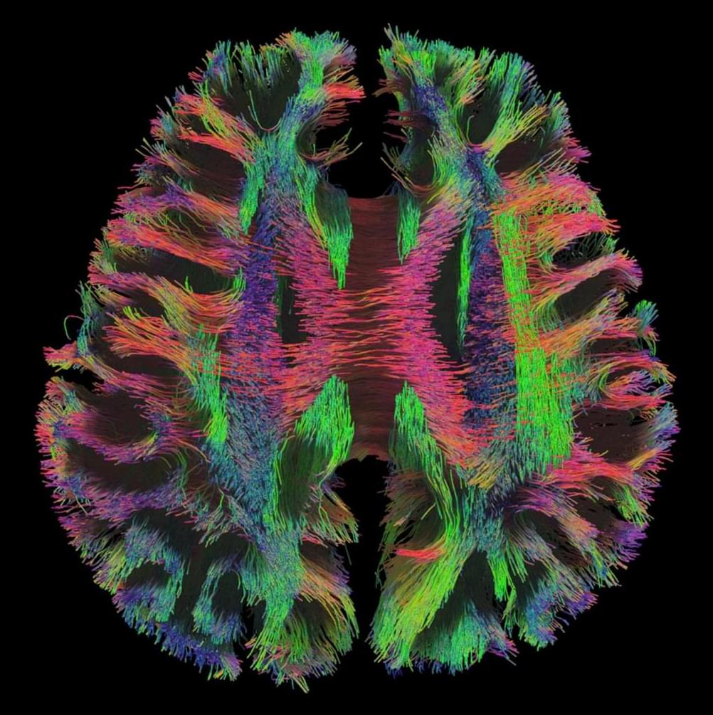
A one-of-a-kind MRI machine helps researchers see the relationship between the structure of the brain and how it functions.
Scientists know that colorectal cancer cells require large amounts of iron and that as cancer becomes more aggressive, the cells have even higher amounts of iron. Normal cells with high levels of iron would undergo a type of iron-related cell death called ferroptosis. But in cancer cells, the iron continues to accumulate well beyond normal levels without succumbing to expected cell death processes.
Researchers from the University of Michigan Health Rogel Cancer Center have now discovered a key metabolic pathway that allows colorectal cancer cells to accumulate large quantities of iron. Blocking that pathway reduced iron levels and caused the cancer cells to die.
In this new study, published in Cell Metabolism, researchers started by looking at the known pathways involved in ferroptosis, assuming something in this process was awry. But knocking out these typical ferroptotic enzymes had no impact on tumor growth. So they dug deeper into mitochondrial metabolism.
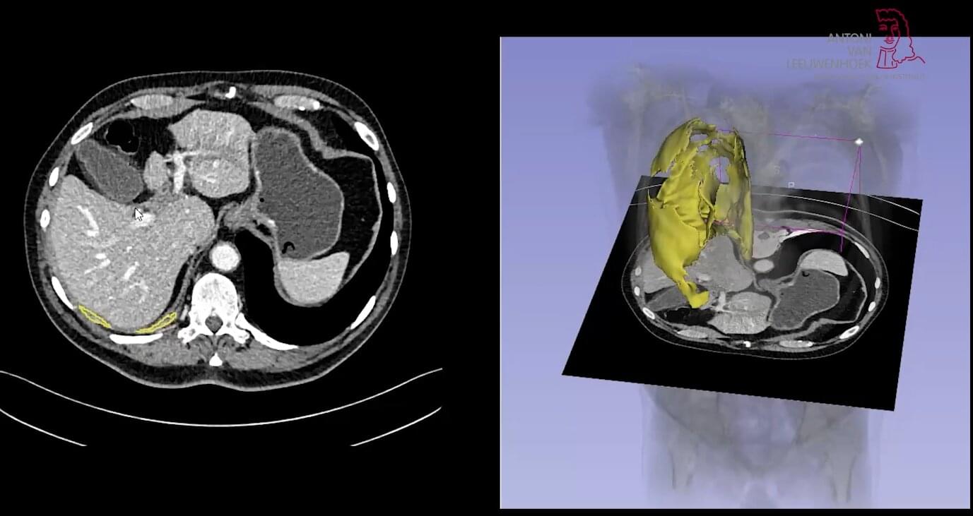
Physicians and researchers at the Netherlands Cancer Institute have developed an AI model that outperforms physicians in evaluating treatment response in pleural mesothelioma. Far more accurate than the current international standard criteria (RECIST), the model provides patients with greater certainty and tailored treatments. It changes how physicians assess tumors and could accelerate the development of new treatments by making clinical trials more reliable and efficient.
Physicians evaluate treatment response by measuring tumor growth. The current diameter-based RECIST criteria are of limited use for pleural mesothelioma because this cancer type grows in a thin, irregular layer along the lung wall. Where, then, do you measure the diameter to determine whether the therapy is working? This leads to uncertainty and frustration among patients and physicians.
AI experts, radiologists and pulmonologists from the Netherlands Cancer Institute (NKI) have now solved this problem. Together, they developed the AI model ARTIMES, which can measure the entire volume of a tumor and compare it with previous scans.
Thomas M. Jessel, Howard Hughes Medical Institute Investigator, explores the human brain, the sophisticated product of 500 million years of vertebrate evolution, assembled during just nine months of embryonic development. The functions encoded by its trillion nerve cells direct all human behavior. Yet the brain is a biological organ made from the same building blocks as skin, liver and lung. How does the brain acquire its remarkable computational power? Answers lie in the details of its construction — the cellular and molecular mechanisms that drive the formation of thousands of neural circuits, each wired for a specific behavior.
Newborn nerve cells must squeeze through crowded, narrow spaces-through dense tissue, past other cells, between fibers-to reach the areas where they form neural circuits in the brain cortex.
In a new study published in Nature, researchers at Kyoto University’s Institute for Integrated Cell-Material Sciences (WPI-iCeMS) and their collaborators report that this journey causes widespread DNA damage in neurons, resulting in double-strand breaks where both strands of the double helix are completely severed. While this is the most severe type of DNA damage-capable of causing mutations and cell death-the team surprisingly found that it is a normal, routine feature of brain cortex formation, and a healthy brain quickly repairs it before harm occurs.
“The developing brain appears to have evolved to tolerate and repair the neuronal damage efficiently,” says Professor Mineko Kengaku, of WPI-iCeMS, who led the study. “But understanding the limits of that tolerance-and what happens when repair is incomplete-brings us closer to understanding a range of neurological conditions.”
What if quantum information is more fundamental than space, time, matter, or even quantum mechanics itself?
Vlatko Vedral explores the implications of a Q-number-based reality for quantum gravity, pre-Big-Bang cosmology, the nature of time, and the possibility that quantum information lies beneath our deepest physical theories.
1:25 Quantum Gravity and Q Numbers.
4:30 Before the Big Bang.
7:42 Time, the Block Universe, and Q Numbers.
11:56 Quantum Mechanics at All Scales.
14:01 The Next Revolution in Physics.
Vlatko Vedral is a Serbian-born British physicist. He is best known for his contributions to quantum information theory, quantum mechanics, and quantum entanglement. He earned his Bachelor of Science and Doctor of Philosophy degrees from Imperial College London, where he graduated with a PhD.
More from Vlatko Vedral on Closer To Truth:
Closer To Truth: The Podcast: • Closer To Truth: The Podcast.
Closer To Truth contributors: https://closertotruth.com/contributor… to Closer To Truth: / @closertotruthtv Join the Community:
Follow Us:
Closer To Truth, hosted by Robert Lawrence Kuhn and directed by Peter Getzels, presents the world’s greatest thinkers exploring humanity’s deepest questions. Discover fundamental issues of existence. Engage new and diverse ways of thinking. Appreciate intense debates. Share your own opinions. Seek your own answers. #CloserToTruth #Cosmos #VlatkoVedral #QuantumInformation #QuantumGravity.
From ultra-flexible materials redefining brain-computer interfaces (BCIs) to record-shattering global out-licensing deals, China’s biopharmaceutical sector is undergoing a profound qualitative transformation. ShanghaiEye takes you inside the Yunfan Future Factory and the cross-discipline innovation hub hosted by Chia Tai Tianqing (CTTQ)—a subsidiary of top-50 global pharma giant Sino Biopharmaceutical—to explore the cutting-edge ecosystem driving the future of global healthcare.
We examine a breakthrough BCI technology developed in Shanghai: an ultra-flexible photoresist material for neural electrode arrays. Ye Tianyang, CEO and Co-Founder of Yunfan Future, explains how this material—engineered to be 1,000 times softer than the rigid alternatives utilized by Western counterparts like Elon Musk’s Neuralink—exponentially reduces tissue damage and immune rejection. With dozens of human clinical trials already successfully completed worldwide, this innovation highlights the immense strength of Shanghai’s local talent pool and medical device supply chain.
The feature also spotlights the strategic roadmap of China’s pharmaceutical leaders. Eric Tse, CEO of Sino Biopharmaceutical and Chairman of CTTQ, breaks down their vision to build an open, interdisciplinary incubator. This global nexus bridges experts, scholars, and upstream and downstream partners, transforming Shanghai into a premier launchpad for international innovative drugs. Furthermore, Mr. Tse discusses the \.
Brody is professor of neuroscience and molecular biology at Princeton University and a Howard Hughes Medical Institute Investigator. His research focuses is on novel quantitative behaviors that allow exploring high-level cognitive questions using powerful emerging tools for studying neural mechanisms in rodents. Brody’s group uses rats to investigate the neural bases of decision making, working memory, and executive control, using a combination of high-throughput semiautomated behavior as well as computational, electrophysiological, pharmacological and optogenetic methods.
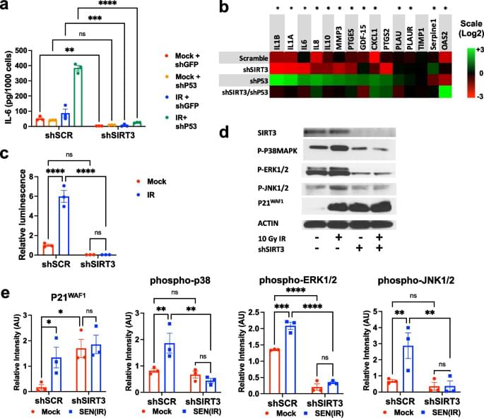
Cellular senescence is a multifaceted stress response marked by stable proliferative arrest and the secretion of diverse biologically active factors, collectively known as the senescence-associated secretory phenotype (SASP). The senescent phenotype is remarkably variable and subject to various regulatory influences. We previously demonstrated that mitochondrial dysfunction induced by diverse stimuli, including the loss of sirtuin 3 (SIRT3), leads to the hyperactivation of AMPK and p53, culminating in senescence while concurrently suppressing much of the proinflammatory SASP. Here, we extend our findings by revealing that the absence of SIRT3 can suppress segments of the SASP even in the absence of p53. Intriguingly, SIRT3 deficiency renders cells resistant to stimulation by exogenous cytokines, such as interleukin-1.