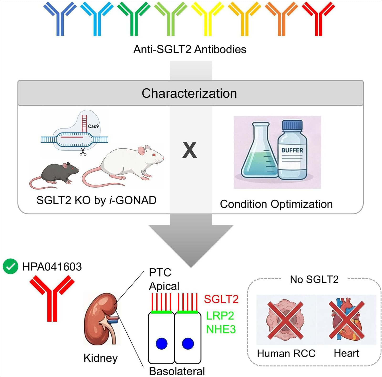
Cambridge researchers discover that gut bacteria can flush PFAS from your body without drugs, neither bloodletting

Understanding of vitamin D physiology is important because about half of the population is being diagnosed with deficiency and treated with supplements. Clinical guidelines were developed based on observational studies showing an association between low serum levels and increased cardiovascular risk. However, new randomized controlled trials have failed to confirm any cardiovascular benefit from supplementation in the general population. A major concern is that excess vitamin D is known to cause calcific vasculopathy and valvulopathy in animal models. For decades, administration of vitamin D has been used in rodents as a reliable experimental model of vascular calcification. Technically, vitamin D is a misnomer. It is not a true vitamin because it can be synthesized endogenously through ultraviolet exposure of the skin. It is a steroid hormone that comes in 3 forms that are sequential metabolites produced by hydroxylases. As a fat-soluble hormone, the vitamin D-hormone metabolites must have special mechanisms for delivery in the aqueous bloodstream. Importantly, endogenously synthesized forms are carried by a binding protein, whereas dietary forms are carried within lipoprotein particles. This may result in distinct biodistributions for sunlight-derived versus supplement-derived vitamin D hormones. Because the cardiovascular effects of vitamin D hormones are not straightforward, both toxic and beneficial effects may result from current recommendations.
To analyze the genome of pancreatic cancer cells, NIST researchers used 13 distinct state-of-the-art whole genome measurement technologies, some of which were only recently developed.
Each method identifies the sequence of DNA nucleotides — adenine (A), cytosine ©, guanine (G) and thymine (T) — in an individual’s genome. However, the methods produce slightly varying results and have different strengths and weaknesses.
NIST’s dataset contains separate results for each of the 13 techniques used to sequence the cancer genome. Scientists performing their analysis can compare their data with NIST’s. If there are discrepancies, they can then determine whether their equipment is working properly and remedy the problem if not.
In the last decade, the incidence of restrictive eating disorders in children, like anorexia-nervosa and avoidant/restrictive food intake disorders (ARFID), has doubled. These disorders have severe consequences for growing children, resulting in nutritional deficiencies and problems with bone development, statural growth and puberty. Most studies have focused on the effects of these disorders in older individuals, and little is currently known about how restrictive eating disorders affect the brain in children or what mechanisms in the brain might be responsible for this restrictive eating behavior.
To get a better understanding of how these early-onset eating disorders work in the brain, researcher Clara Moreau and her team conducted MRI brain scans on 290 children, of which 124 had been hospitalized for early-onset anorexia-nervosa (EO-AN), 50 had been hospitalized for ARFID, and 116 were children with no eating disorders. All participants were under 13 years old, and those who were hospitalized had very low body mass index (BMI) due to restrictive eating. The results were published in Nature Mental Health.
Although EO-AN and AFRID both result in low BMI and malnutrition due to restrictive eating, they are distinct disorders. EO-AN—as well as later onset anorexia-nervosa—is characterized by restrictive eating arising from a distorted body image, while restrictive eating in AFRID arises from sensory issues, such as a dislike of certain food textures, a lack of interest in food or fear of negative health consequences from food. These differences indicate that the disorders probably arise from different mechanisms in the brain.
In a step forward for soft robotics and biomedical devices, Rice University engineers have uncovered a powerful new way to boost the strength and durability of silicone-based soft devices without changing the materials themselves. Their study, published in a special issue of Science Advances, focuses on printed and musculoskeletal robotics and offers a predictive framework that connects silicone curing conditions with adhesion strength, enabling dramatic improvements in performance for both molded and 3D-printed elastomer components.
New research into mitotic chromosomes has found that they are covered in a liquid-like coating that could allow them to bounce off one another, like bumper cars, protecting them from being damaged during cell division.
The research from Dr. Daniel Booth and Professor Amanda Wright at the University of Nottingham, in partnership with the universities of Glasgow and Kent, looked into the coatings of mitotic chromosomes, the highly condensed and organized structures that DNA morphs into during cell division. Their findings are published in the journal Nature Communications.
The mitotic chromosome periphery (MCP) is a poorly understood “coat” that covers all chromosomes. Previous work from Dr. Booth revealed that Ki-67, an important cancer biomarker, organizes the entire coat and when the coat is removed chromosomes become sticky and clump together, meaning that cells are sometimes too sick to divide properly. This raised the possibility that the MCP might have undiscovered specialist biophysical properties.
Two cannabis-derived compounds have shown remarkable effectiveness against fungal pathogens in laboratory tests, according to new Macquarie University research.
Promise: Dr Hue Dinh, pictured, and colleagues at the Macquarie University Galleria laboratory are hopeful their discovery will lead to new treatments for common skin infections.
In a study published in The Journal of Neglected Tropical Diseases (PLOS NTDs), researchers discovered that bioactives Cannabidiol (CBD) and Cannabidivarin (CBDV) killed harmful Cryptococcus neoformans - a WHO-listed priority fungal pathogen. The compounds also killed dermatophytes that cause common skin infections, and much faster than existing treatments.
All day long, our brains carry out complicated and energy-intensive tasks such as remembering, solving problems, and making decisions.
To supply the energy these tasks require while conserving this precious fuel, the brain has evolved a system that allows it to quickly and efficiently send blood only to the areas that need it most in any given moment. This system is essential to brain function and overall health, yet how it works has remained somewhat of a mystery.
Now, a team led by researchers at Harvard Medical School has uncovered new details of how the brain moves blood to active areas in real time. Their findings are published July 16 in Cell.
In experiments in mice, the team discovered that the brain uses specialized channels in the lining of its blood vessels to communicate where blood is needed.
“This work helps us understand how you can get that super-important blood supply to the correct areas of the brain on a time scale that is useful,” said co-lead author Luke Kaplan, a research fellow in neurobiology in the Blavatnik Institute at HMS.
If confirmed in additional studies in animals and humans, the findings could be used to better understand findings on brain imaging tests such as functional MRI (fMRI). The insights may also advance understanding of neurodegenerative diseases, in which this communication system often breaks down, leading to cognitive problems.
Precision and personalized medicine for disease management necessitates real-time, continuous monitoring of biomarkers and therapeutic drugs to adjust treatment regimens based on individual patient responses. This study introduces a wearable Microneedle-based Continuous Biomarker/Drug Monitoring (MCBM) system, designed for the simultaneous, in vivo pharmacokinetic and pharmacodynamic evaluation for diabetes. Utilizing a dual-sensor microneedle and a layer-by-layer nanoenzyme immobilization strategy, the MCBM system achieves high sensitivity and specificity in measuring glucose and metformin concentrations in skin interstitial fluid (ISF). Seamless integration with a smartphone application enables real-time data analysis and feedback, fostering a pharmacologically informed approach to diabetes management. The MCBM system’s validation and in vivo trials demonstrate its precise monitoring of glucose and metformin, offering a tool for personalized treatment adjustments. Its proven biocompatibility and safety suit long-term usage. This system advances personalized diabetes care, highlighting the move towards wearables that adjust drug dosages in real-time, enhancing precision and personalized medicine.
Real-time monitoring of drugs and biomarkers is essential for personalized diabetes care. Here, the authors present a wearable microneedle sensor system enabling simultaneous in vivo monitoring of glucose and metformin in interstitial fluids for personalized medicine.