Toggle light / dark theme

The Revolving Door of Adenovirus Cell Entry: Not All Pathways Are Equal

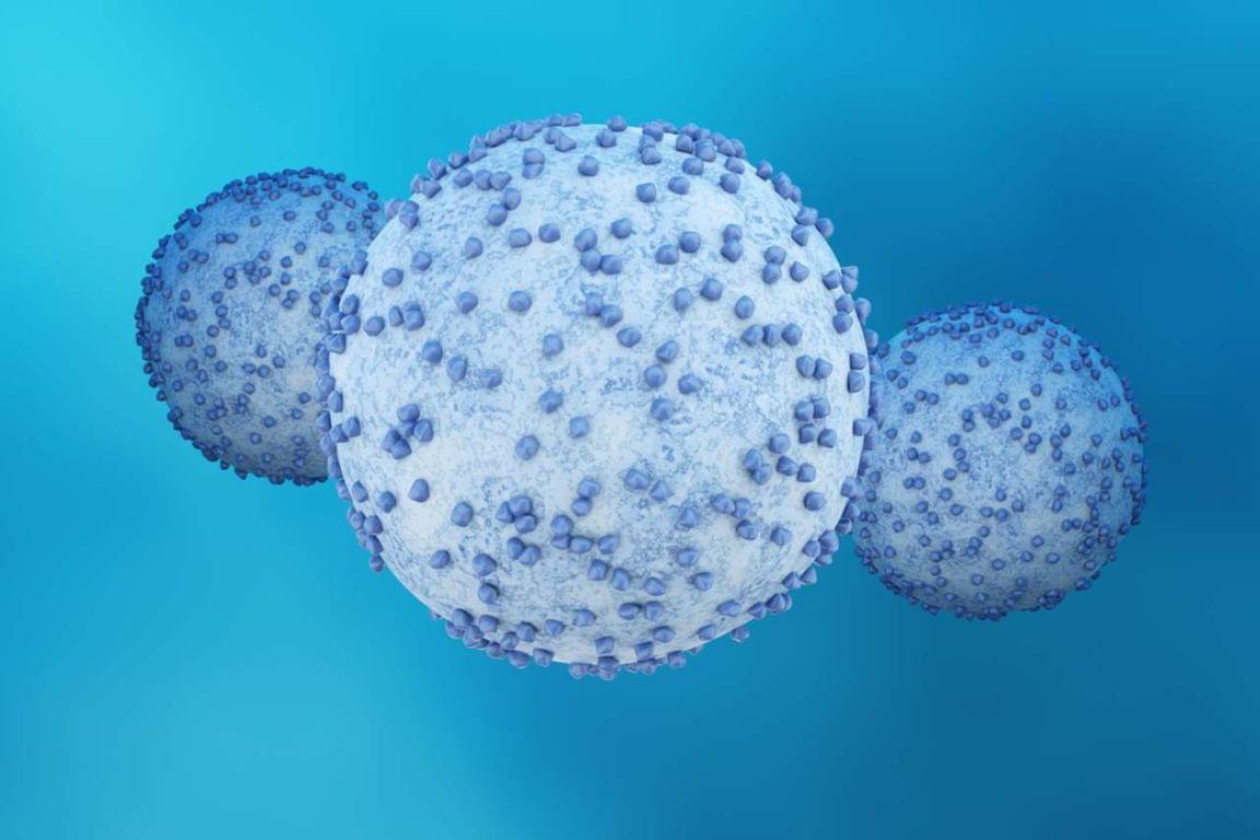
An interesting review on adenoviral cell entry and trafficking. Its discussion of how species B adenoviruses tolerate lower endosomal pH and accumulate in later-endosomal compartments before escaping were particularly intriguing. Link.


Adenoviruses represent exceptional candidates for wide-ranging therapeutic applications, from vectors for gene therapy to oncolytics for cancer treatments. The first ever commercial gene therapy medicine was based on a recombinant adenovirus vector, while most recently, adenoviral vectors have proven critical as vaccine platforms in effectively controlling the global coronavirus pandemic. Here, we discuss factors involved in adenovirus cell binding, entry, and trafficking; how they influence efficiency of adenovirus-based vectors; and how they can be manipulated to enhance efficacy of genetically modified adenoviral variants. We focus particularly on endocytosis and how different adenovirus serotypes employ different endocytic pathways to gain cell entry, and thus, have different intracellular trafficking pathways that subsequently trigger different host antiviral responses.

Opposing protein pathways steer skin stem cells toward renewal or repair

Two proteins with opposing functions orchestrate the development and maintenance of healthy skin, Stanford Medicine researchers have found. Modulating their activity with topical drugs could reduce inflammation, aid wound healing and slow or halt the growth of skin cancer, the researchers believe. The findings are published in the journal Science.

The proteins are part of a family called ubiquitin-like proteins. Ubiquitination controls the targeted destruction and disposal of unneeded proteins in a cell. But in the skin, certain ubiquitin-like proteins instead switch on or off wide swaths of genes involved in cellular growth and development, the study found. In particular, they trigger progenitor (stem) cells in the lower layer of the skin to either mature and migrate to the skin surface or to self-renew.

“These two ubiquitin-like protein systems are remarkably dedicated and opposite in their functions,” said Paul Khavari, MD, Ph.D., chair of dermatology at the Stanford School of Medicine and senior author of the study. “One promotes the stem-cell state while the other drives differentiation. It’s like having two opposing forces that determine a cell’s fate.”

Iron accumulation in the brain may contribute to neurodegeneration

Neurodegenerative diseases affect tens of millions of people worldwide. Among these, Alzheimer’s and Parkinson’s diseases are the most common; in the United States alone, the Alzheimer’s Disease Association and Parkinson’s Foundation report roughly 7 million people with Alzheimer’s and another million with Parkinson’s. An intriguing clue lies in the tangled mystery of neurodegeneration that scientists are working to solve: iron accumulation.

Scientists have noticed that iron can slowly build up inside neurons. Early in life, this iron accumulation appears to have little effect on neuronal function. However, later in life, it can contribute to a slow neuronal demise. Salk Institute researchers studied nerve cells to figure out whether and how this iron accumulation relates to neurodegenerative diseases. They found that the excess iron stuck in neurons lowers the cells’ defenses, making them more vulnerable to stressors and other cellular insults through a process they named chronoferroptosis.

The study, published in Cell Death Discovery on June 18, 2026, points to iron accumulation as a key target in the effort to predict, prevent and treat neurodegenerative diseases.

First 3D views of human cone opsins reveal how daylight vision reacts so fast

The retina of the human eye contains 6–7 million cone cells. These cells contain light-sensitive proteins known as cone opsins. They enable us to perceive our surroundings in detail in daylight. They allow us to see the world in thousands of colors: red strawberries, green leaves, the blue sky. They also enable us to see all the objects around us clearly. And they allow us to perceive fast movements, such as the rush of a train or the flight of a dragonfly.

Often, however, these all-rounders of daylight vision are also involved in retinal diseases. Impairment of cone receptor function, caused by genetic mutations or other degenerative processes, can lead to disorders such as color blindness and age-related macular degeneration (AMD), a disease affecting the central retina and causing progressive vision loss.

In a new study, Polina Isaikina and Sarah L. Schmidt, two researchers from the Center for Life Sciences at PSI, have succeeded for the first time in determining the three-dimensional structure of human cone opsins in their dark state and showing how their molecular architecture enables their rapid activation by light.

Proteomic analysis of dental enamel from 20 Homo naledi individuals shows no male markers

The morphological homogeneity of the middle Pleistocene hominin Homo naledi is reflected in its dental proteome, with no evidence supporting confident identification of male individuals, and the detection of both derived and ancestral amino acid substitutions in Homo naledi.

Preventing the next pandemic using AI-designed vaccines

For most of human history, infectious diseases were the main causes of morbidity and mortality. Advances in sanitation, antibiotics, vaccines, and public health dramatically shifted that balance, particularly in high-income countries, where life expectancy has increased by nearly 40 years over the past century. Yet the COVID-19 pandemic provided a stark reminder that infectious threats can still reshape societies almost overnight. Between 2019 and 2021 alone, life expectancy in the US fell by more than two years, and recent modelling suggests there is roughly a 50 percent chance of another COVID-scale pandemic occurring within the next 25 years.

Historically, the vaccine development model has been largely reactive and variant-driven, but the industry is now actively shifting toward proactive and universal vaccinology to get ahead of evolving pathogens. Recent results from a first-in-human clinical trial led by the University of Cambridge and its spin-out DIOSynVax, published in the Journal of Infection, provide early clinical evidence of this shift, demonstrating the safety of an AI-designed “super-antigen” intended to provide broad viral coverage.

Teaching AI to Invent Enzymes Nature Never Imagined

Evolution is an extraordinary engine for enzymatic diversity, yet the chemistry it has explored remains a narrow slice of what DNA can encode. Deep generative models can design new proteins that bind ligands, but none have created enzymes without pre-specifying catalytic residues.

In this webinar, Chenghao Liu and Jarrid Brooks from the Arnold Lab at Caltech will introduce DISCO (DIffusion for Sequence-structure CO-design). This multimodal model co-designs protein sequence and 3D structure around arbitrary biomolecules, as well as inference-time scaling methods that optimize objectives across both modalities. Conditioned solely on reactive intermediates, DISCO designs diverse heme enzymes with novel active-site geometries. These enzymes catalyze new-to-nature carbene-transfer reactions, including alkene cyclopropanation, spirocyclopropanation, B-H, and C(sp^3)-H insertions, with high activities exceeding those of engineered enzymes. Random mutagenesis of a selected design further confirmed that enzyme activity can be improved through directed evolution. By providing a scalable route to evolvable enzymes, DISCO broadens the potential scope of genetically encodable transformations.

Sleep deprivation increases levels of the synaptic density marker SV2A in the human brain

The synaptic homeostasis hypothesis posits that sleep is essential for restoring cerebral equilibrium by downscaling synaptic connections that progressively strengthen and accumulate metabolic costs during wakefulness. While previously supported only by preclinical animal models, a recent study provides direct in vivo evidence of this mechanism in humans. Researchers evaluated 40 volunteers, half of whom underwent 28 hours of continuous sleep deprivation, utilizing Positron Emission Tomography (PET) to quantify levels of the SV2A protein, a reliable biomarker for synaptic density. The findings revealed that prolonged wakefulness significantly elevated SV2A levels across multiple brain regions, most notably in the hippocampus and thalamus. Furthermore, during a subsequent two-hour recovery sleep period, these elevated SV2A levels were strongly correlated with enhanced slow-wave activity, a primary electrophysiological marker of deep sleep and homeostatic sleep pressure. These results validate the synaptic homeostasis hypothesis in humans, demonstrating a measurable biological link between sleep deprivation, the accumulation of neural connections, and the restorative drive for deep, slow-wave sleep.


The synaptic homeostasis hypothesis (SHY) [14] posits that wakefulness promotes synaptic potentiation due to environmental interactions and learning [5]. The strengthening of connections during waking elevates energy consumption, results in the accumulation of proteins and receptors that compete for the limited anatomical space in the skull and diminishes the signal-to-noise ratios in the neuronal network, ultimately saturating the capacity for learning. Sleep allows for synaptic down-selection, preserving energy and network efficiency. While the SHY has been supported by anatomical and molecular studies in animals, human evidence has remained limited due to the invasive nature of most techniques for quantifying synaptic strength.

Studies in animals indicate that anatomical or molecular markers of synaptic strength increase during wake and decline during sleep [6]. Firing rates in rodents indicate increased cortical excitability during wakefulness and decreased cortical excitability during sleep. In humans, cortical excitability is an indirect measure of plasticity. Findings from studies using transcranial magnetic stimulation (TMS) translated the findings from the above-mentioned rodent studies (reviewed in [7]). However, some in-vitro and in-vivo studies of synaptic strength in animals reveal opposite results, which may be due to differences in the type of marker, examined brain regions, cortical layers, or housing of animals (reviewed in [8]).

Synaptic vesicle glycoprotein 2A (SV2A) [9] is an integral membrane protein located on synaptic vesicles. Recent advances in PET imaging with tracers such as [¹⁸F]SynVesT-1 enable the noninvasive measurement of SV2A binding in the living human brain [10,11], allowing new opportunities to examine state-dependent synaptic changes. However, whether this reflects presynaptic terminal number, vesicle complement, SV2A expression per vesicle, or excitatory/inhibitory-synapse composition cannot be resolved with in vivo imaging. While SV2A availability is commonly interpreted as a proxy measure of synaptic density, we refer to it here as ‘SV2A-indexed synaptic density’ to reflect this interpretation while acknowledging its underlying biological ambiguity.

Sugar-coated nanoparticles show promise for treating most aggressive form of brain cancer

Researchers at Oregon State University have potentially found a new way to treat the most aggressive form of brain cancer, glioblastoma, whose two-year survival rate is less than 30%.

The study, led by Oleh Taratula, Olena Taratula and Yoon Tae Goo of the OSU College of Pharmacy, addresses what they describe as the two most persistent obstacles to effective glioblastoma treatment: delivering therapeutic agents through the blood-brain barrier, the cell network that acts as a security checkpoint between the bloodstream and the central nervous system, and then getting those agents to preferentially target tumors.

In research published in the Journal of Controlled Release, the scientists demonstrate the novel treatment technique in a mouse model. They loaded lipid nanoparticles with genetic material that promotes tumor suppression, then coated the nanoparticles with a type of sugar. The result was a 50% median increase in glioblastoma survival time.

/* */