Toggle light / dark theme

Tiny molecular rings open new ways for designing better medicines

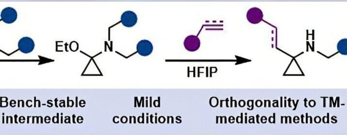
Cyclopropanes are organic chemistry’s smallest rings. Three carbon atoms are joined in a triangle, creating a compact and unusually strained structure. Despite—or partly because of—this unusual geometry, cyclopropanes are found in many biologically active natural products and have important applications in medicines and drug discovery, from antidepressants to antibiotics and antiviral research.

Connect these carbon triangles to an amine, a functional group with a nitrogen at its center, and you get aminocyclopropanes. They are of particular interest in pharmaceutical research because they can be used to replace another substructure commonly found in drug molecules: α,α-gem-dimethylamines (compounds in which the aminocyclopropane’s triangle is “opened” by disconnecting one edge).

Such molecular substitutes that resemble an existing part of a drug but can alter important properties, such as biological activity and metabolic stability, are referred to as bioisosteres and have shown beneficial effects in countless cases.

Untangling the meta-biomaterial puzzle one property at a time

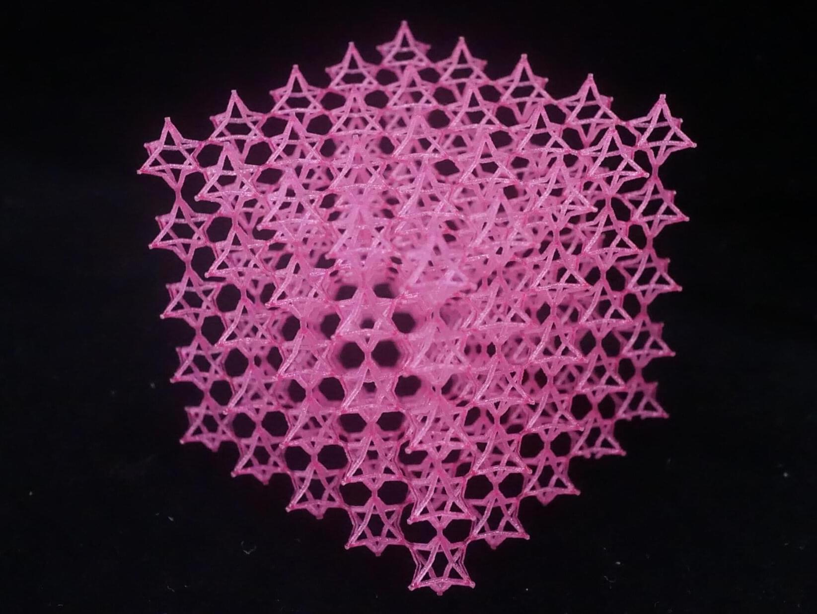
From repairing damaged tissues to developing better implants, many medical developments depend on materials that can mimic the complex properties of human tissue. Meta-biomaterials are among the most promising candidates. By tailoring their geometry, researchers can create materials with properties similar to those of natural tissues. But there is a catch: Changing one property often changes several others at the same time. TU Delft scientists have now developed a method to decouple these properties.

Their work, published in Nature Communications, helps researchers understand how individual material properties influence cell behavior and could accelerate the development of next-generation biomaterials.

Meta-biomaterials are engineered materials whose properties are determined not by their chemical composition but by their internal architecture. This allows researchers to design structures with carefully tuned mechanical, morphological and mass-transport properties. Such control is particularly valuable in biomedical engineering, where materials need to do more than simply replace damaged tissue. They must also interact with cells and actively support tissue regeneration.

Depression in Later Life May Be an Early Warning Sign of Alzheimer’s

Depression in later life may be an early warning from the brain, appearing years before Alzheimer’s begins to affect memory and thinking

A new study in JNeurosci found that faster accumulation of tau, a protein closely tied to Alzheimer’s, was associated with worsening depressive symptoms in older adults who remained cognitively healthy. Teodora Markova of Brandeis University and her colleagues investigated whether changes in mood might track the gradual buildup of tau in the aging brain.

Tau normally helps support the internal structure of neurons. When it becomes abnormal, however, it can collect into tangles that interfere with brain function. These tangles are one of the defining biological features of Alzheimer’s disease, alongside deposits of amyloid beta.

Super-resolution imaging reveals details inside living cells

A new fluorescence microscopy technique that generates super-resolution images from a single camera exposure could help researchers study rapid movements within cells that are difficult to capture with existing methods.

Super-resolution microscopy has transformed cell biology by allowing scientists to see cellular structures and details that are too small to be imaged clearly with conventional light microscopes. Some methods achieve super-resolution by combining information from hundreds or thousands of image frames taken over time.

However, this works best when biological samples like cells or tissues have been fixed, or preserved with chemicals. Otherwise, structures like mitochondria, microtubules and DNA—which are constantly moving, changing and interacting—appear blurry using existing super-resolution techniques.

Lentiviral In Vivo CD19 CAR TCell Therapy in Neurologic Autoimmune Disorders

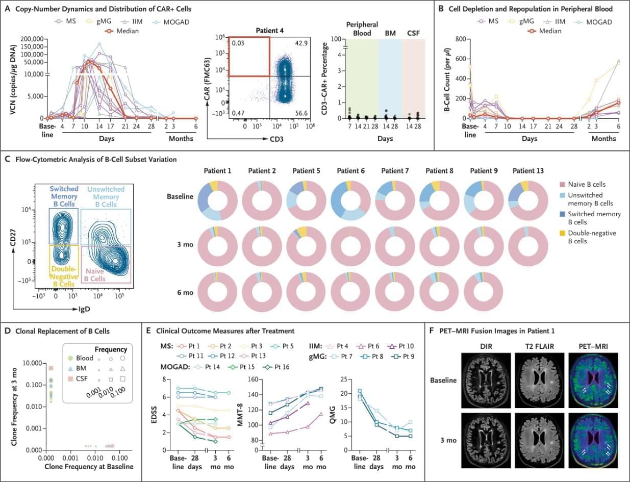
An international team of scientists has successfully tested an innovative in vivo CAR-T therapy, JY231, capable of treating severe autoimmune diseases with a single intravenous infusion. Published in the New England Journal of Medicine, the experimental treatment uses a harmless genetically modified viral vector to deliver genetic instructions directly into a patient’s T-lymphocytes, reprogramming them to target and destroy defective B-cells that produce autoantibodies. In a clinical trial involving 16 patients with treatment-resistant conditions such as multiple sclerosis, myasthenia gravis, and myopathy, the therapy demonstrated over 99% accuracy. Within two months, patients’ bone marrow began producing healthy immune cells, and after six months of observation, participants exhibited significant clinical improvements, including the elimination of chronic fatigue and the partial restoration of cognitive and muscular functions. Researchers are now preparing for large-scale randomized trials to definitively confirm the method’s long-term safety and efficacy.


Among 16 patients with refractory neurologic autoimmune disorders, lentiviral CD19 CAR T-cell therapy was associated with manageable side effects, complete B-cell depletion, and preliminary clinical improvement across disease groups.

Plasmalogens Eliminate AgingAssociated Synaptic Defects and MicrogliaMediated Neuroinflammation in Mice

Neurodegeneration is a pathological condition in which nervous system or neuron losses its structure, function, or both leading to progressive neural degeneration. Growing evidence strongly suggests that reduction of plasmalogens (Pls), one of the key brain lipids, might be associated with multiple neurodegenerative diseases, including Alzheimer’s disease (AD). Plasmalogens are abundant members of ether-phospholipids. Approximately 1 in 5 phospholipids are plasmalogens in human tissue where they are particularly enriched in brain, heart and immune cells. In this study, we employed a scheme of 2-months Pls intragastric administration to aged female C57BL/6J mice, starting at the age of 16 months old. Noticeably, the aged Pls-fed mice exhibited a better cognitive performance, thicker and glossier body hair in appearance than that of aged control mice. The transmission electron microscopic (TEM) data showed that 2-months Pls supplementations surprisingly alleviate age-associated hippocampal synaptic loss and also promote synaptogenesis and synaptic vesicles formation in aged murine brain. Further RNA-sequencing, immunoblotting and immunofluorescence analyses confirmed that plasmalogens remarkably enhanced both the synaptic plasticity and neurogenesis in aged murine hippocampus. In addition, we have demonstrated that Pls treatment inhibited the age-related microglia activation and attenuated the neuroinflammation in the murine brain. These findings suggest for the first time that Pls administration might be a potential intervention strategy for halting neurodegeneration and promoting neuroregeneration.

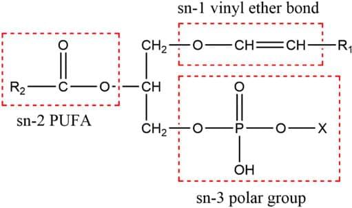
Plasmalogens (Pls) are a special type of vinyl ether-bonded phospholipids actively participating in structure and function of biological membranes. Approximately 20% of phospholipids are plasmalogens in human tissue, where they are particularly rich in the brain, heart, and immune cells (Lessig and Fuchs 2009; Braverman and Moser 2012). In brain, ethanolamine plasmalogens (PlsEtns) constitute approximately 60 and 80% of the total ethanolamine phospholipids in gray and white matter, respectively (Macala et al., 1983). Pls are also concentrated in specialized membranes, such as sarcolemma, myelin, and synaptic vesicles (Post et al., 1988; Takamori et al., 2006; Poitelon et al., 2020). Reduced levels of PlsEtns have been found to be associated with aging (Pradas et al., 2019) and several neurodegenerative diseases, including Alzheimer’s disease (AD) (Guan et al., 1999; Han et al., 2001; Goodenowe et al., 2007; Wood 2010; Wood et al., 2015; Yamashita et al.
Super-resolution imaging reveals that cohesin prevents local mixing of compact, active genome domains

The human genome is about two meters (6.6 feet) long, yet it is folded inside a cell nucleus only about 10 micrometers in diameter. To fit into this tiny space, DNA is wrapped around histone proteins to form nucleosomes, which are further organized into chromatin. For decades, chromatin has often been described in two simple forms: euchromatin, which is active, open and accessible, and heterochromatin, which is more compact and repressed.

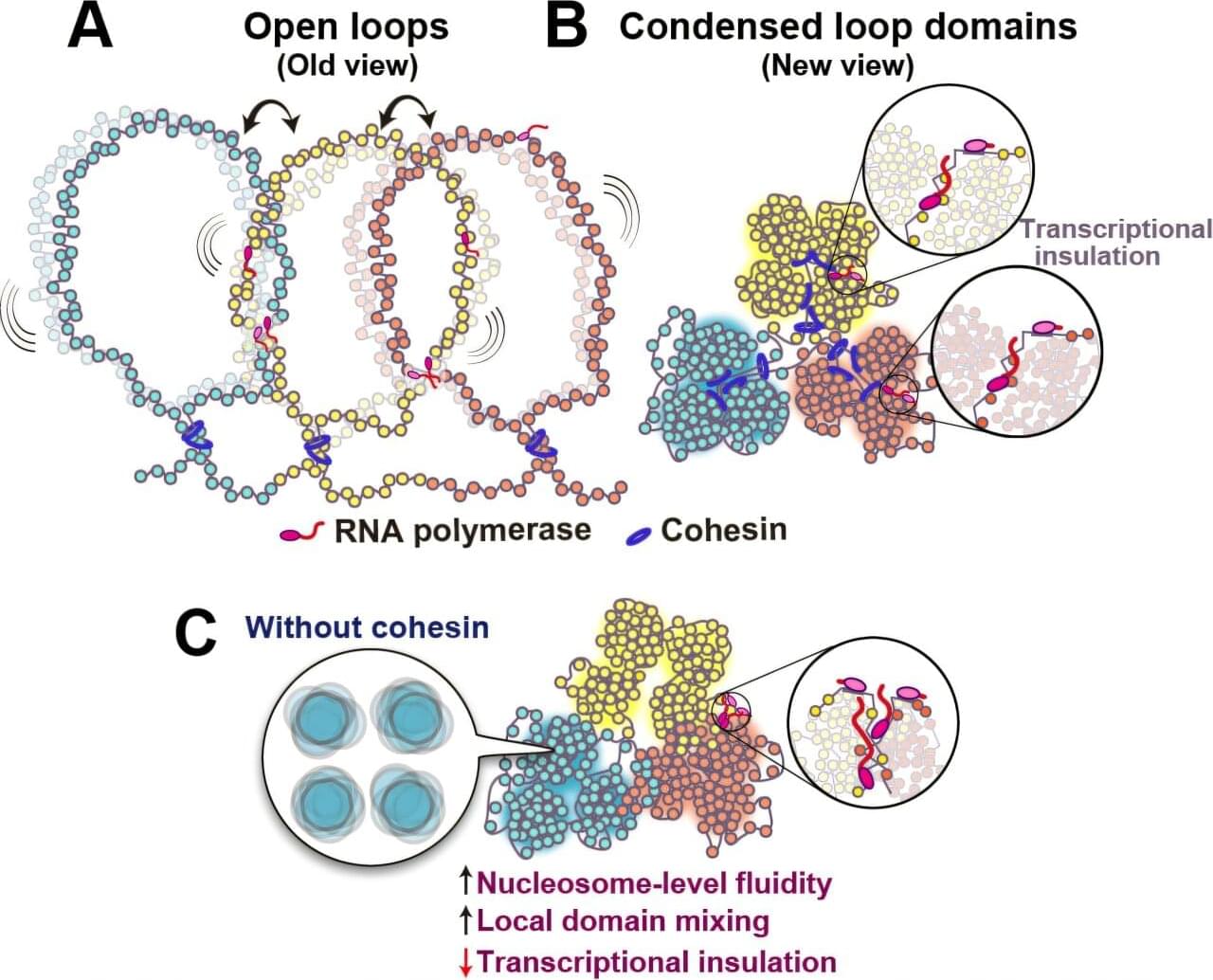
However, a new study from an international team led by Kazuhiro Maeshima, a professor at the National Institute of Genetics, ROIS (Research Organization of Information and Systems) and SOKENDAI, has challenged this simple textbook view. The researchers demonstrated that euchromatin in living human cells is not merely open and loose but forms dynamic condensed domains. This domain organization helps prevent the mixing of neighboring domains.

The team further found that cohesin, a ring-shaped protein complex best known for organizing genome architecture, prevents local mixing between these condensed euchromatic domains for proper gene regulation in living human cells. The study was published in Nature Genetics on Sept. 8, 2026.

Scientists discover a hidden immune signal that helps spinal cords regrow

A surprising immune system signal may help explain why zebrafish can regenerate damaged spinal cords so effectively. Researchers found that certain neutrophils release Il-4, which calms harmful inflammation and allows injured nerve fibers to grow again. Without these cells, healing stalled, but adding Il-4 restored regeneration. Scientists now want to know whether the same mechanism could someday be harnessed to improve spinal cord repair in humans.

New insights on CRISPR/Cas9based therapy for breast Cancer Environment

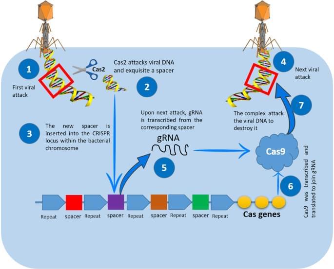
CRISPR/Cas9 has revolutionized genome-editing techniques in various biological fields including human cancer research. Cancer is a multi-step process that encompasses the accumulation of mutations that result in the hallmark of the malignant state. The goal of cancer research is to identify these mutations and correlate them with the underlying tumorigenic process. Using CRISPR/Cas9 tool, specific mutations responsible for cancer initiation and/or progression could be corrected at least in animal models as a first step towards translational applications. In the present article, we review various novel strategies that employed CRISPR/Cas9 to treat breast cancer in both in vitro and in vivo systems.

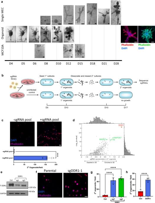
Breast tissue regeneration is driven by cellmatrix interactions coordinating multilineage stem cell differentiation through DDR1 Communications

Breast tissue regeneration is driven by cell-matrix interactions coordinating multi-lineage stem cell differentiation through DDR1.


Mammary morphogenesis is a complex process. Here the authors describe how stem cells build a three-dimensional self-organizing multi-lineage tissue by showing that positional signals from the extracellular matrix through the collagen receptor DDR1 lead stem cells to differentiate into multi-lineage committed multi-layered progeny.

/* */