Python infostealers are spreading from Windows to macOS via Google Ads, ClickFix lures, and fake installers to steal credentials and financial data.


Microsoft has started rolling out built-in Sysmon functionality to some Windows 11 systems enrolled in the Windows Insider program.
Microsoft first revealed plans to integrate Sysmon natively into Windows 11 and Windows Server in November, when it also confirmed that it will soon release detailed documentation.
Sysmon (short for System Monitor) is a free Microsoft Sysinternals tool (and a Windows system service and device driver) that monitors for and blocks malicious/suspicious activity, logging it to the Windows Event Log.
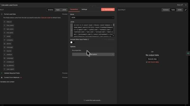
Multiple critical vulnerabilities in the popular n8n open-source workflow automation platform allow escaping the confines of the environment and taking complete control of the host server.
Collectively tracked as CVE-2026–25049, the issues can be exploited by any authenticated user who can create or edit workflows on the platform to perform unrestricted remote code execution on the n8n server.
Researchers at several cybersecurity companies reported the problems, which stem from n8n’s sanitization mechanism and bypass the patch for CVE-2025–68613, another critical flaw addressed on December 20.

The U.S. Cybersecurity and Infrastructure Security Agency (CISA) ordered government agencies to patch their systems against a five-year-old GitLab vulnerability that is actively being exploited in attacks.
GitLab patched this server-side request forgery (SSRF) flaw (tracked as CVE-2021–39935) in December 2021, saying it could allow unauthenticated attackers with no privileges to access the CI Lint API, which is used to simulate pipelines and validate CI/CD configurations.
“When user registration is limited, external users that aren’t developers shouldn’t have access to the CI Lint API,” the company said at the time.


A new threat actor called Amaranth Dragon, linked to APT41 state-sponsored Chinese operations, exploited the CVE-2025–8088 vulnerability in WinRAR in espionage attacks on government and law enforcement agencies.
The hackers combined legitimate tools with the custom Amaranth Loader to deliver encrypted payloads from command-and-control (C2) servers behind Cloudflare infrastructure, for more accurate targeting and increased stealth.
According to researchers at cybersecurity company Check Point, Amaranth Dragon targeted organizations in Singapore, Thailand, Indonesia, Cambodia, Laos, and the Philippines.