The light-controlled electron current could open new paths for sensing, telecommunications, and other advanced technologies.
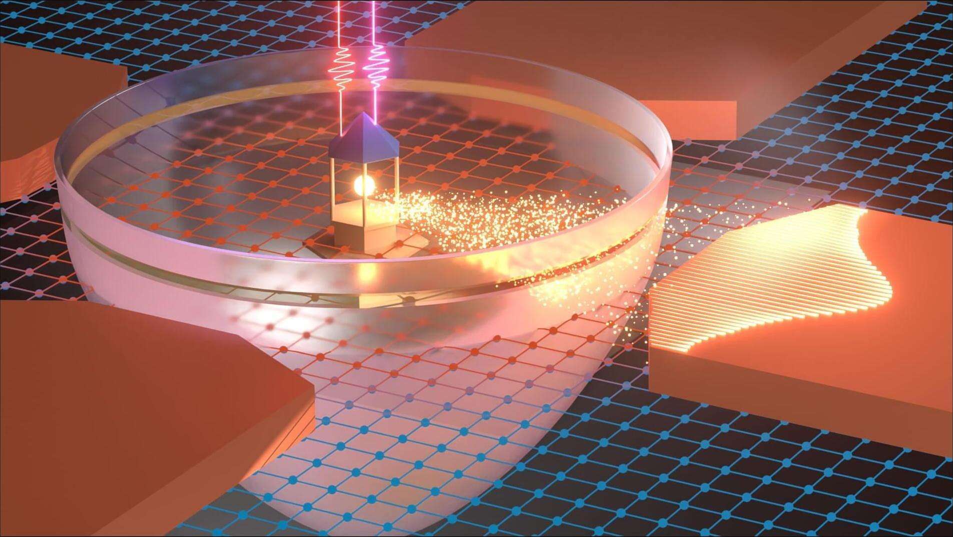
A pair of laser beams can now send electrons through a semiconductor in a chosen direction without any external electrical power. Researchers at the University of Michigan built the device to explore a previously unobserved physical effect and demonstrate that light alone can both generate and steer an electronic current.
The work could eventually support technologies that combine optics and electronics, including sensing, imaging, and telecommunications. By improving how signals move within and between devices, the effect may also allow those signals to carry more information.