Toggle light / dark theme

Brain circuit needed to incorporate new information may be linked to schizophrenia

One of the symptoms of schizophrenia is difficulty incorporating new information about the world. This can lead people with schizophrenia to struggle with making decisions and, eventually, to lose touch with reality.

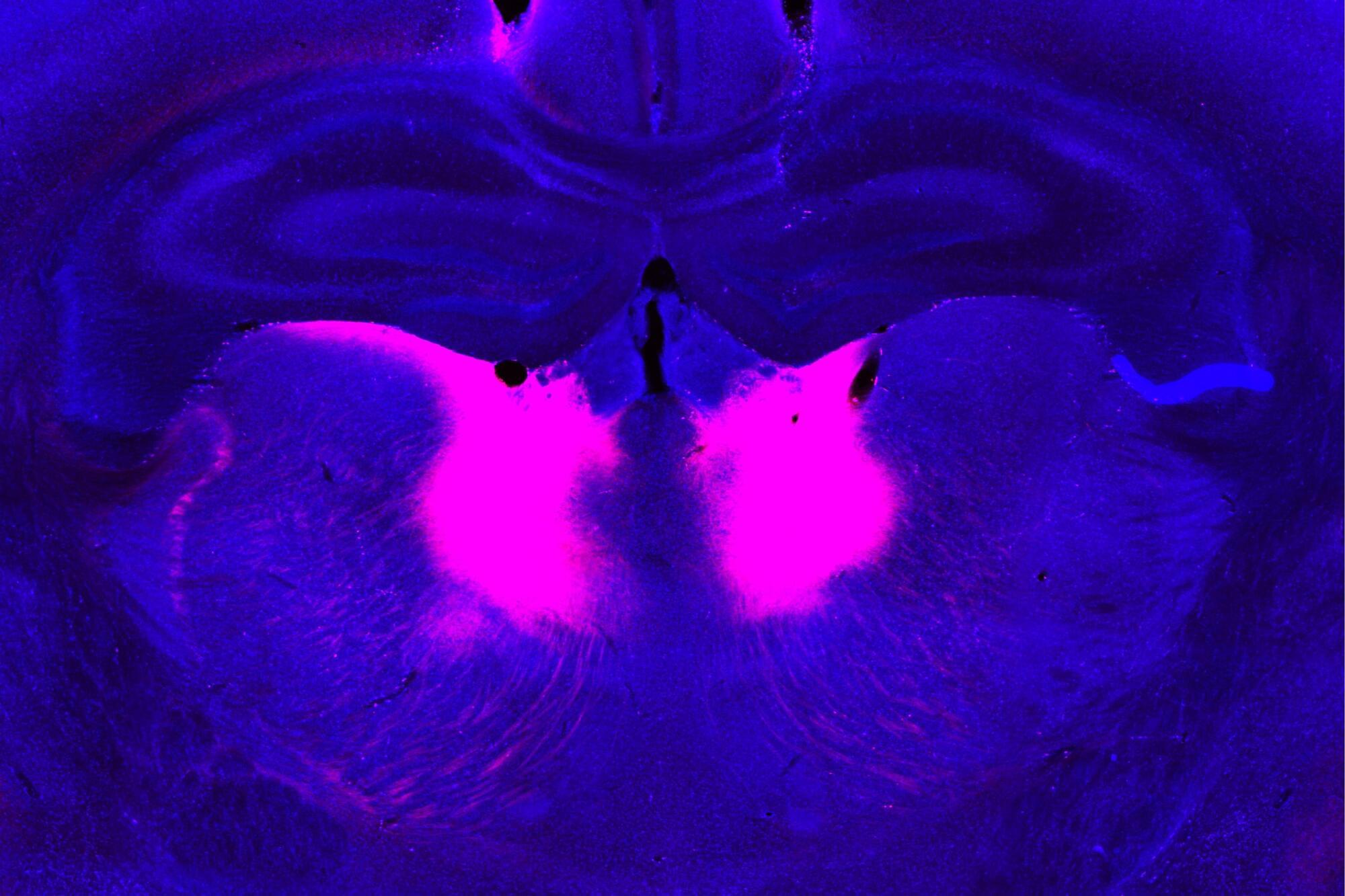
MIT neuroscientists have now identified a gene mutation that appears to give rise to this type of difficulty. In a study of mice, the researchers found that the mutated gene impairs the function of a brain circuit that is responsible for updating beliefs based on new input.

This mutation, in a gene called grin2a, was originally identified in a large-scale screen of patients with schizophrenia. The new study suggests that drugs targeting this brain circuit could help with some of the cognitive impairments seen in people with schizophrenia.

Brain keeps familiar routes intact as new experiences get layered on top, study suggests

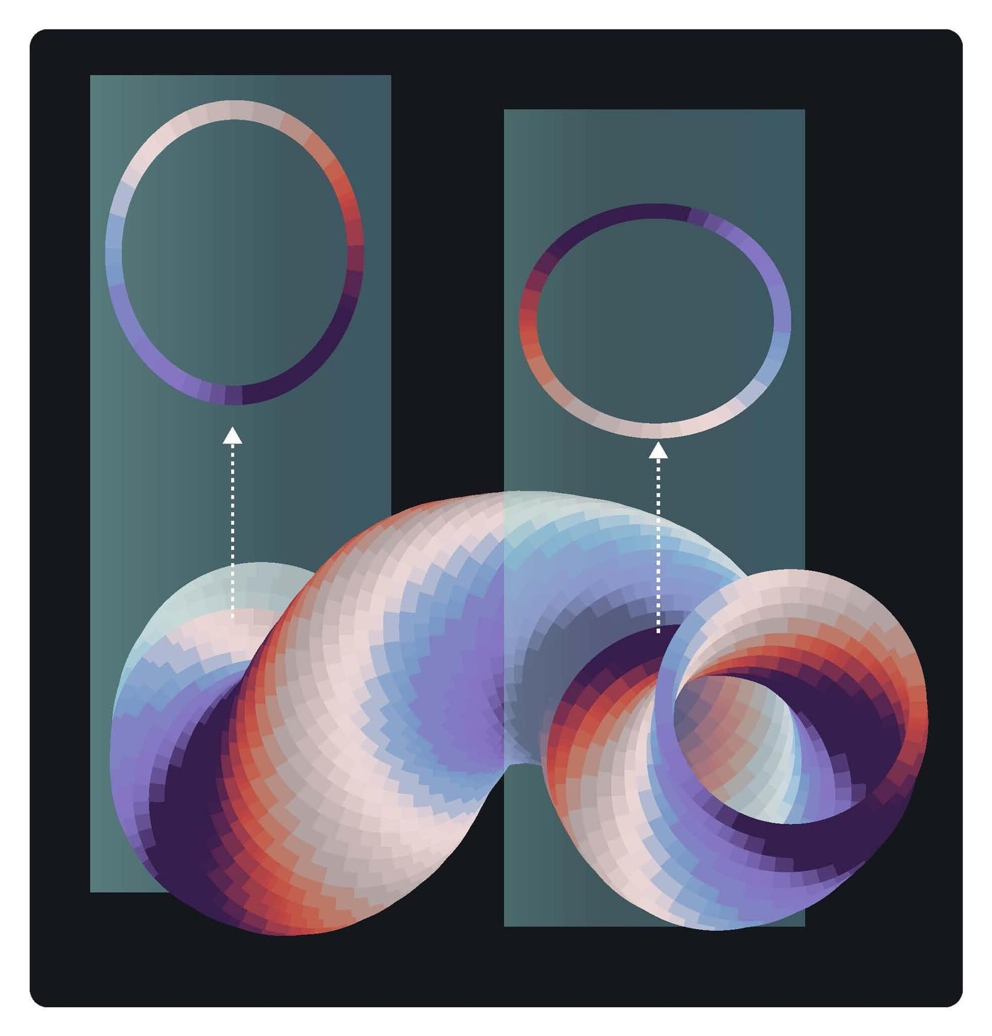
Every time we move through a familiar environment, the hippocampus consults an internal map, a detailed spatial representation built up through repeated experience. But what happens when something unexpected occurs on a well-known route? Researchers at the University Hospital Bonn (UKB) and the University of Bonn demonstrated in a mouse model that the brain does not redraw its maps from scratch. Instead, it annotates them, preserving the underlying spatial layout while overlaying new information on top of the existing map. The paper is published in the Proceedings of the National Academy of Sciences.

The hippocampus, the brain’s working memory, is shaped like a seahorse and is located in the temporal lobe of both the left and right hemispheres. Hippocampal CA3 circuits, which link information and support the recognition of memories, keep their spatial maps stable while layering new annotations on top, much like a navigation app that preserves your route while flagging an incident ahead.

A Bonn-based research team arrived at these findings by recording the activity of CA3 axons in mice traversing a familiar linear running route. At a fixed point along the route, the scientists introduced a mildly aversive but harmless air puff stimulus, comparable to an unexpected obstacle on a road, and tracked how the hippocampal network updated its representation before, during and after the event.

Scientists Revived Activity in a Disembodied Human Brain

Further Reading.

Thumbnail image credit: Not alive, but not dead… FEATURED SCIENCE ARTICLE.
MRI image: Britannica: brain.
anatomy.

Not alive, but not dead: disembodied human brains used for drug testing.
https://www.science.org/content/artic…

Restoration of brain circulation and cellular functions hours.
https://pubmed.ncbi.nlm.nih.gov/30996…

Science #explained #brains #organoid #sciencenews

Frontiers: For nearly a decade, the idea that ‘the body keeps the score’ has shaped public and clinical understanding of trauma (van der Kolk, 2014)

It is an enticing metaphor—implying that experience is literally inscribed in flesh, that the body bears the scars of what the mind cannot face. Yet recent advances in computational and systems neuroscience reveal that this image, while emotionally compelling, is biologically inaccurate. The body proper does not store trauma; the brain dynamically reenacts it through maladaptive inference. What endures after trauma is not a memory lodged in non-innervated tissue but a collapse of flexibility—a loss of metastability, the brain’s ability to fluidly switch among semi-stable network states.

In computational terms, trauma over-weights the precision of danger priors: the brain assigns excessive confidence to threat predictions, constraining inference based on the prior premise of enduring danger. The result is hypervigilance, flashbacks, and avoidance—symptoms of a system caught in self-confirming predictions. Mathematically, this overconfidence means one cannot escape local minima—in a free energy landscape—that become deeply and precisely engrained (i.e., trapped in a ravine with steep sides, where precision corresponds to the local curvature or steepness).

This rigidity contrasts with a healthy brain’s metastable dynamics, where neuronal networks continually integrate and segregate in response to context. This allows neuronal dynamics to explore multiple (unstable) interpretations of the world. Hellyer and colleagues demonstrated that metastability is a hallmark of cognitive flexibility: the capacity for neural coalitions to assemble transiently and adapt quickly. Using both empirical and computational approaches, Hellyer et al. (2015) showed that reduced metastability arising from damage to the structural connectome was associated with diminished cognitive flexibility and impaired information processing. Trauma erodes this fluidity, trapping the brain in narrow basins of fear and defensive salience. To restore mental health is not about ‘releasing’ stored emotion but reestablishing dynamic equilibrium enabling the brain’s ability to move with graceful agility over a landscape of beliefs, commitments and intentions.