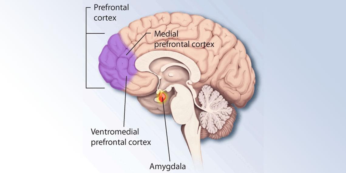
Toggle light / dark theme

For nearly his entire life, Dr. Stuart Hameroff has been fascinated with the bedeviling question of consciousness. But instead of studying neurology or another field commonly associated with the inner workings of the brain, it was Hameroff’s familiarity with anesthetics, a family of drugs that famously induces the opposite of consciousness, that fueled his curiosity.

“I thought about neurology, psychology, and neurosurgery, but none of those… eemed to be dealing with the problem of consciousness,” says Hameroff, a now-retired professor of anesthesiology from the University of Arizona. Hameroff recalls a particularly eye-opening moment when he first arrived at the university and met the chairman of the anesthesia department. “He says ‘hey, if you want to understand consciousness, figure out how anesthesia works because we don’t have a clue.’”

Hameroff’s work in anesthesia showed that unconsciousness occurred due to some effect on microtubules and wondered if perhaps these structures somehow played a role in forming consciousness. So instead of using the neuron, or the brain’s nerve cells, as the “base unit” of consciousness, Hameroff’s ideas delved deeper and looked at the billions of individual tubulins inside microtubules themselves. He quickly became obsessed.

Researchers at University of California San Diego have developed and tested a new software package, called Spatial Modeling Algorithms for Reactions and Transport (SMART), that can realistically simulate cell-signaling networks—the complex systems of molecular interactions that allow cells to respond to diverse cues from their environment.

Cell-signaling networks involve many distinct steps and are also greatly influenced by the complex, three-dimensional shapes of cells and subcellular components, making them difficult to simulate with existing tools. SMART offers a solution to this problem, which could help accelerate research in fields across the life sciences, such as , pharmacology and .

The researchers successfully tested the new software in biological systems at several different scales, from cell signaling in response to adhesive cues, to calcium release events in subcellular regions of neurons and , to the production of ATP (the energy currency in cells) within a detailed representation of a single mitochondrion.

“Engineering” sleeping consciousness could reduce nightmares, treat insomnia—and even be induce specific dreams just for fun.

By Michelle Carr edited by Mark Fischetti

I routinely control my own dreams. During a recent episode, in my dream laboratory, my experience went like this: I was asleep on a twin mattress in the dark lab room, wrapped in a cozy duvet and a blanket of silence. But I felt like I was awake. The sensation of being watched hung over me. Experimenters two rooms over peered at me through an infrared camera mounted on the wall. Electrodes on my scalp sent them signals about my brain waves. I opened my eyes—at least I thought I did—and sighed. Little specks of pink dust hovered in front of me. I examined them curiously. “Oh,” I then thought, realizing I was asleep, “this is a dream.”

People with breathing problems during sleep may have a larger hippocampus, the area of the brain responsible for memory and thinking, according to a study published in the December 18, 2024, online issue of Neurology.

The study, which included mostly Latino people, also found that those with lower during sleep had changes in the deep parts of the brain, the , a common finding of decreased that develops with age.

Sleep disordered breathing is a range of conditions that cause abnormal breathing during sleep, including snoring and . Obstructive sleep apnea is when a person stops breathing five or more times per hour. When breathing stops, it can lower oxygen levels, affecting the brain.

Researchers from Max Planck Florida Institute for Neuroscience have discovered a new pathway to forming long-term memories in the brain. Their work, published in Nature Neuroscience, suggests that long-term memory can form independently of short-term memory, a finding that opens exciting possibilities for understanding memory-related conditions.

Our brain works diligently to record our experiences into memories, creating representations of our daily events that stay with us for short time periods. Current scientific theories of memory formation suggest that short-term memories are stored in what we can imagine as a temporary art exhibition in our brain before being cleared out for representations of new experiences.

A tiny fraction of these short-term memories—those most relevant to us—are moved to a more permanent exhibit, our long-term memory, where they are stored for days, years, or decades.

The brain is sometimes called the most complex machine in the known universe. But the thoughts that it outputs putter along at a trifling 10 bits per second, the pace of a conversation.

By Rachel Nuwer

People tend to have the sense that their inner thoughts and feelings are much richer than what they are capable of expressing in real time. Elon Musk has spoken publicly about this “bandwidth problem,” as he described it to podcaster Joe Rogan. Musk is so bothered by this, in fact, that he has made it one of his long-term goals to create an interface that allows the human brain to communicate directly with a computer, unencumbered by the slow speed of speaking or writing.

PHOENIX — Mayo Clinic announces the results of an innovative treatment approach that may offer improvement in overall survival in older patients with newly diagnosed glioblastoma while maintaining quality of life. Glioblastoma is the most lethal type of primary brain cancer due to its aggressive nature and its treatment-resistant characteristics. It is the most common form of primary brain cancer. Each year an estimated 14,500 people in the U.S. are diagnosed with the disease. Results of Mayo Clinic’s phase 2, single-arm study are published in The Lancet Oncology.

Sujay Vora, M.D., radiation oncologist at Mayo Clinic, led a team of researchers investigating the use of short-course hypofractionated proton beam therapy incorporating advanced imaging techniques in patients over the age of 65 with newly diagnosed World Health Organization (WHO) grade 4, malignant glioblastoma.

Results showed that 56% of participants were alive after 12 months and the median overall survival was 13.1 months.” As compared to prior phase 3 studies in an older population having a median survival of only six to nine months, these results are promising,” says Dr. Vora. “In some cases, patients with tumors that have favorable genetics lived even longer, with a median survival of 22 months. We are very excited about these results.”

Microgravity is known to alter the muscles, bones, the immune system and cognition, but little is known about its specific impact on the brain. To discover how brain cells respond to microgravity, Scripps Research scientists, in collaboration with the New York Stem Cell Foundation, sent tiny clumps of stem-cell derived brain cells called “organoids” to the International Space Station (ISS).

Surprisingly, the organoids were still healthy when they returned from orbit a month later, but the cells had matured faster compared to identical organoids grown on Earth—they were closer to becoming adult neurons and were beginning to show signs of specialization. The results, which could shed light on potential neurological effects of space travel, appear in Stem Cells Translational Medicine.

“The fact that these cells survived in space was a big surprise,” says co-senior author Jeanne Loring, Ph.D., professor emeritus in the Department of Molecular Medicine and founding director of the Center for Regenerative Medicine at Scripps Research. “This lays the groundwork for future experiments in space, in which we can include other parts of the brain that are affected by neurodegenerative disease.”