Every thought, memory, and feeling we experience depends on trillions of tiny connection points in the brain called synapses. These are the junctions where one neuron passes signals to another, forming the vast communication network known as the connectome—the brain’s wiring diagram. Although scientists have developed powerful tools to increase or decrease neural activity, directly redesigning the brain’s physical wiring has remained far more difficult.
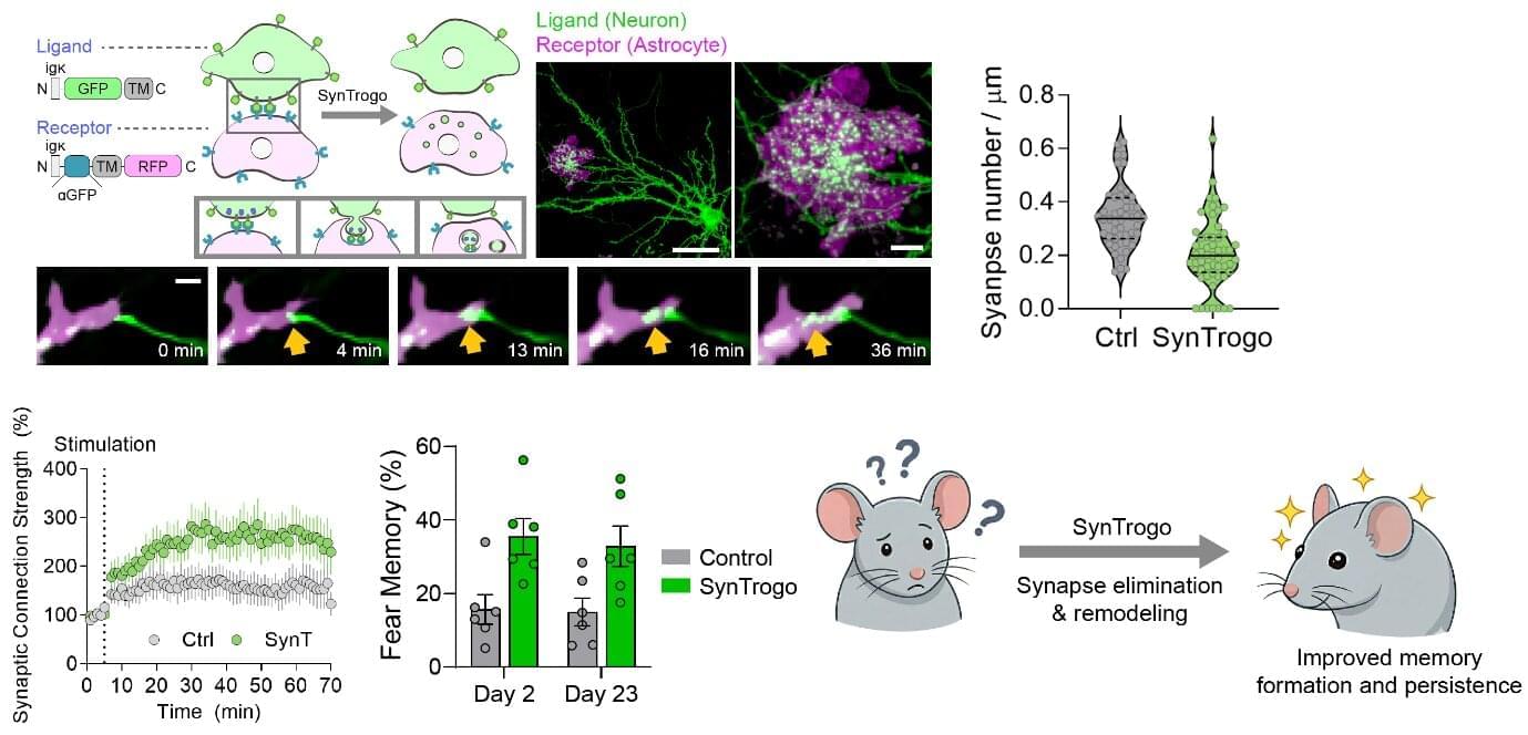
A research team led by Dr. Sangkyu Lee and Director C. Justin Lee at the Center for Memory and Glioscience within the Institute for Basic Science (IBS), in collaboration with Dr. Kea Joo Lee of the Korea Brain Research Institute (KBRI), has now developed a molecular tool that makes such structural editing possible. The new platform, called SynTrogo (Synthetic Trogocytosis), enables researchers to induce astrocytes to selectively remodel synaptic connections in a targeted brain circuit. The paper is published in the journal Nature Communications.
The brain already has a natural mechanism for refining its wiring. During development and throughout life, unneeded or weak connections are removed in a process known as synaptic pruning, much like trimming unnecessary branches from a tree. This pruning is partly carried out by astrocytes—star-shaped glial cells that closely surround synapses and help maintain the neural environment. When this process becomes dysregulated, either through too much or too little pruning, it has been linked to disorders such as schizophrenia, autism spectrum disorder, and Alzheimer’s disease.

