Humans are musical animals 4 million years in the making, explained by music expert Michael Spitzer.

Summary: The psychedelic compound DMT (Dimethyltryptamine) increases connectivity across the brain, allowing for greater communication between different areas and networks. The brain changes are most prominent in brain areas linked to higher functioning, such as imagination.
Source: Imperial College London.
Scientists have gleaned new insights into how psychedelics alter conscious experience via their action on brain activity.
Recently biologists discovered how to generate new neurons in the adult brain. This is an incredible breakthrough that has enormous potential to revolutionize neurodegenerative disease research. By generating genetically-mutated mice with a unique gene that activates dormant neural stem cells, scientists were able to generate new neurons in the brain. For years, scientists have been searching for ways to promote the growth of new neurons in the brain, especially in individuals with neurodegenerative diseases such as Alzheimer’s and Parkinson’s. This new discovery could lead to new treatments and therapies that could help to restore brain function and improve the quality of life for millions of people around the world.
Leslie Samuel, founder of Interactive Biology, gives some context for the importance of genetic trading between organisms for scientific research, and notes how the loss of nerve cells in the brain is one of the hallmarks of neurodegenerative diseases. The ability to generate new neurons in the adult brain could be a game-changer in the field of neurology.
Leslie’s Thoughts

Julian Jaynes was living out of a couple of suitcases in a Princeton dorm in the early 1970s. He must have been an odd sight there among the undergraduates, some of whom knew him as a lecturer who taught psychology, holding forth in a deep baritone voice. He was in his early 50s, a fairly heavy drinker, untenured, and apparently uninterested in tenure. His position was marginal. “I don’t think the university was paying him on a regular basis,” recalls Roy Baumeister, then a student at Princeton and today a professor of psychology at Florida State University. But among the youthful inhabitants of the dorm, Jaynes was working on his masterpiece, and had been for years.
From the age of 6, Jaynes had been transfixed by the singularity of conscious experience. Gazing at a yellow forsythia flower, he’d wondered how he could be sure that others saw the same yellow as he did. As a young man, serving three years in a Pennsylvania prison for declining to support the war effort, he watched a worm in the grass of the prison yard one spring, wondering what separated the unthinking earth from the worm and the worm from himself. It was the kind of question that dogged him for the rest of his life, and the book he was working on would grip a generation beginning to ask themselves similar questions.
The Origin of Consciousness in the Breakdown of the Bicameral Mind, when it finally came out in 1976, did not look like a best-seller. But sell it did. It was reviewed in science magazines and psychology journals, Time, The New York Times, and the Los Angeles Times. It was nominated for a National Book Award in 1978. New editions continued to come out, as Jaynes went on the lecture circuit. Jaynes died of a stroke in 1997; his book lived on. In 2000, another new edition hit the shelves. It continues to sell today.
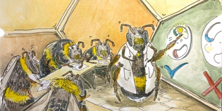
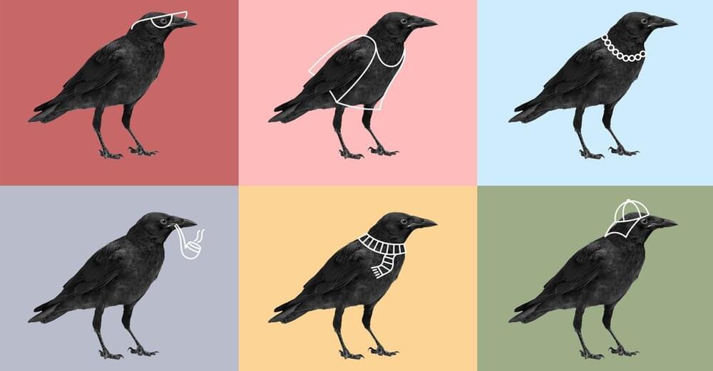
Social insects like bees demonstrate a remarkable range of behaviors, from working together to build structurally complex nests (complete with built-in climate control) to the pragmatic division of labor within their communities. Biologists have traditionally viewed these behaviors as pre-programmed responses that evolved over generations in response to external factors. But two papers last week reported results indicating that social learning might also play a role.
The first, published in the journal PLoS Biology, demonstrated that bumblebees could learn to solve simple puzzles by watching more experienced peers. The second, published in the journal Science, reported evidence for similar social learning in how honeybees learn to perform their trademark “waggle dance” to tell other bees in their colony where to find food or other resources. Taken together, both studies add to a growing body of evidence of a kind of “culture” among social insects like bees.
“Culture can be broadly defined as behaviors that are acquired through social learning and are maintained in a population over time, and essentially serves as a ‘second form of inheritance,’ but most studies have been conducted on species with relatively large brains: primates, cetaceans, and passerine birds,” said co-author Alice Bridges, a graduate student at Queen Mary University of London who works in the lab of co-author Lars Chittka. “I wanted to study bumblebees in particular because they are perfect models for social learning experiments. They have previously been shown to be able to learn really complex, novel, non-natural behaviors such as string-pulling both individually and socially.”
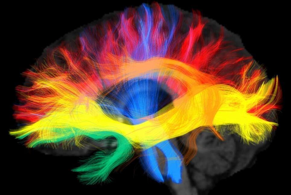
Scientists at the Max Planck Institute for Human Cognitive and Brain Sciences in Leipzig have found evidence that the language we speak shapes the connectivity in our brains that may underlie the way we think. With the help of magnetic resonance tomography, they looked deep into the brains of native German and Arabic speakers and discovered differences in the wiring of the language regions in the brain.
Xuehu Wei, who is a doctoral student in the research team around Alfred Anwander and Angela Friederici, compared the brain scans of 94 native speakers of two very different languages and showed that the language we grow up with modulates the wiring in the brain. Two groups of native speakers of German and Arabic respectively were scanned in a magnetic resonance imaging (MRI) machine.
The high-resolution images not only show the anatomy of the brain, but also allow to derive the connectivity between the brain areas using a technique called diffusion-weighted imaging. The data showed that the axonal white matter connections of the language network adapt to the processing demands and difficulties of the mother tongue.
Two newly discovered genes have been linked to schizophrenia while a previously known gene associated with schizophrenia risk has also been linked to autism in a massive new study.
Scientists say the findings increase our understanding of brain diseases and could lead to new treatment targets.
Importantly, this is the first known investigation to look at the risk of schizophrenia in different groups of people, especially those with African ancestry. It revealed rare harmful variations in gene proteins raise the risk of schizophrenia in all ethnic groups.