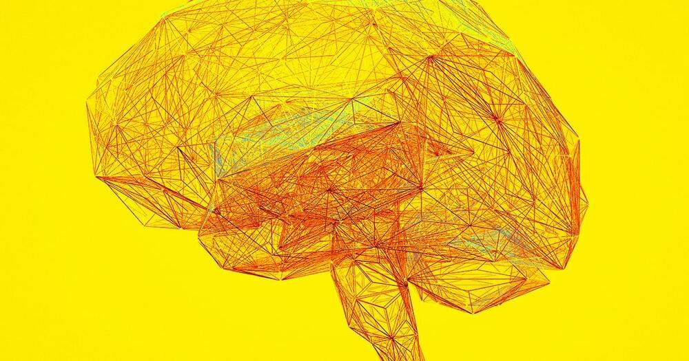
A pair of philosophers sounded the alarm on the dystopian applications they see being ushered in by brain-computer interface (BCI) technology.
“This interface could revolutionize the way we interact with technology.”
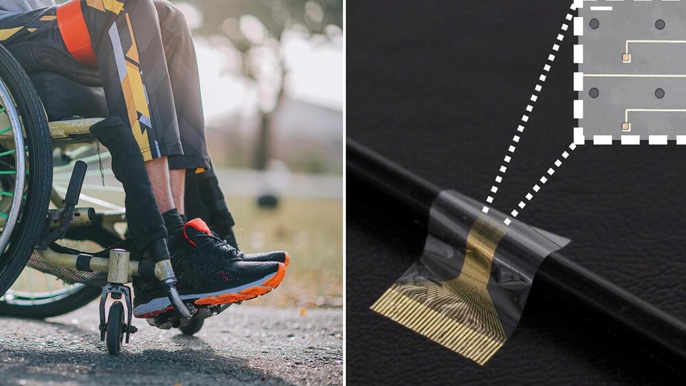
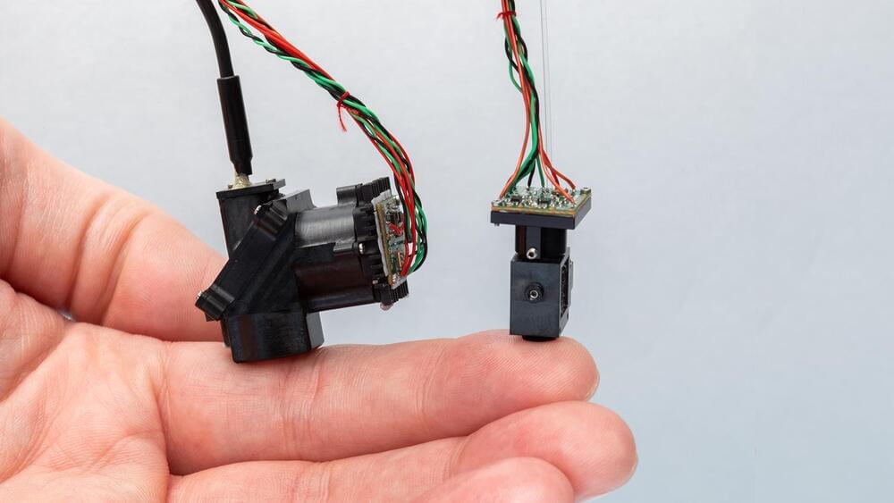
Researchers from the University of Cambridge have created a new type of neural implant that could restore limb function in paralyzed limbs.
There have been former attempts at using neural implants to restore limb function, but these mostly failed. This is because scar tissue can envelop the electrodes over time, disrupting the connection between the device and the nerve.
University of Cambridge.
The developed device works in sync between the brain and paralyzed limbs — it combines flexible electronics and human stem cells to “better integrate” with the nerve and drive limb function, according to a press release.
Near Death Experience Research Foundation the largest collection of Near Death Experiences (NDE) in over 23 Languages. With thousands of full-text near death experiences posted. Share your near death experience, research, spiritually transforming events, consciousness studies, extensive information and research.
In this video, we will explore the positional system of the brain — hippocampal place cells. We will see how it relates to contextual memory and mapping of more abstract features.
OUTLINE:
00:00 Introduction.
00:53 Hippocampus.
1:27 Discovery of place cells.
2:56 3D navigation.
3:51 Role of place cells.
4:11 Virtual reality experiment.
7:47 Remapping.
11:17 Mapping of non-spatial dimension.
13:36 Conclusion.
_____________
REFERENCES:
1) Anderson, M.I., Jeffery, K.J., 2003. Heterogeneous Modulation of Place Cell Firing by Changes in Context. J. Neurosci. 23, 8827–8835. https://doi.org/10.1523/JNEUROSCI.23-26-08827.
2) Aronov, D., Nevers, R., Tank, D.W., 2017. Mapping of a non-spatial dimension by the hippocampal–entorhinal circuit. Nature 543719–722. https://doi.org/10.1038/nature21692
3) Bostock, E., Muller, R.U., Kubie, J.L., 1991. Experience-dependent modifications of hippocampal place cell firing. Hippocampus 1193-205. https://doi.org/10.1002/hipo.
For individuals suffering from drug addiction, certain cues—whether it’s specific people, places or things—can trigger powerful cravings for repeated use.
A new University of Michigan study has identified brain signals, traditionally associated with inflammation, contributing to people’s vulnerability to addiction. With repeated drug use with the same exposure to cues, some individuals develop an inability to control their drug use, even in the face of negative consequences.
The study is published in the journal eNeuro.
The study authors claim their microscope can provide colored real-time images of hard-to-reach parts of the spinal cord that couldn’t be accessed previously.
Pain is a powerful feeling but have you ever wondered how pain works on a cellular level? Well, a team of scientists at the San Diego-based Salk Insitute has actually figured out a way to see the internal neural mechanism associated with pain.
In their recently published study, they propose wearable microscopes using which they were able to check how nerve cells in the spinal cord of mice process pain signals.
Salk Institute.
http://www.ted.com Philosopher Dan Dennett makes a compelling argument that not only don’t we understand our own consciousness, but that half the time our brains are actively fooling us.
TEDTalks is a daily video podcast of the best talks and performances from the TED Conference, where the world’s leading thinkers and doers are invited to give the talk of their lives in 18 minutes. TED stands for Technology, Entertainment, and Design, and TEDTalks cover these topics as well as science, business, politics and the arts. Watch the Top 10 TEDTalks on TED.com, at http://www.ted.com/index.php/talks/top10
Daniel Dennett.
May 14, 2014
Serious thinkers contend that free will cannot exist in a deterministic universe — one in which events are the singular outcomes of the conditions in which they occur. The alternative view, that free will is prerequisite for personal responsibility and morality, is the basis of our legal and religious institutions. Philosopher Daniel Dennett unravels this conundrum and asks whether we must jettison one of these notions, or whether they can co-exist. He then asks: if free will is an illusion, as many scientists say, should we conclude that we don’t need real free will to be responsible for our actions?
Daniel Dennett is the Austin B. Fletcher Professor of Philosophy and Director, Center for Cognitive Studies at Tufts University.
Derk Pereboom claims that free will is impossible because of its incompatibility with both determinism and indeterminism. Also he defends a robust nonreductive physicalism. It says that although consciousness can’t be reduced to physical it’s not something over and above physical.
The interview was taken by Vadim Vasiliev and Dmitry Volkov. Below you’ll see a list of questions of the interview.
1. The most influential books.
2. What are the differences between notions of moral responsibility and basic desert?
3. Which type of punishment should be eliminated if we find out that there is no justification for basic desert?
4. Is indignation as a reaction on wrongdoing a kind of irrational emotion?
5. How was the manipulation argument invented?
6. Why you’ve recently changed a presentation of the first case of Manipulation Argument?
7. How does the problem of free will relate to the problem of mental causation?
8. Could the problem of personal identity pose difficulties for moral responsibility and for basic desert? And why causal determinism is at the focus of free will debate?
9. Is there a real difference between hard incompatibilist’s position and that of compatibilists?
10. Can you list or name some differences and similarities between you and Daniel Dennett?
11. Could cognitive science and neuroscience eliminate the discussion on free will?
12. What is a definition of mental?
13. What were the most important changes of your views?
14. What is meaning of life?
15. What is your current research?