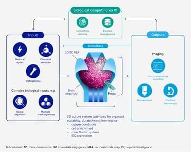
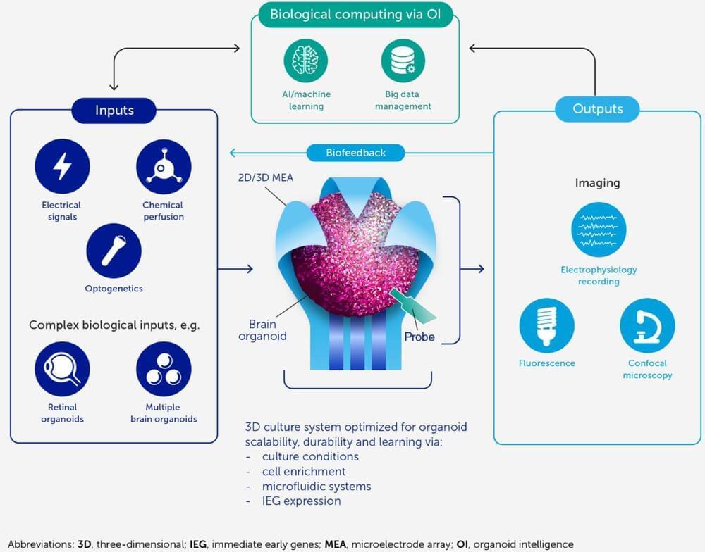
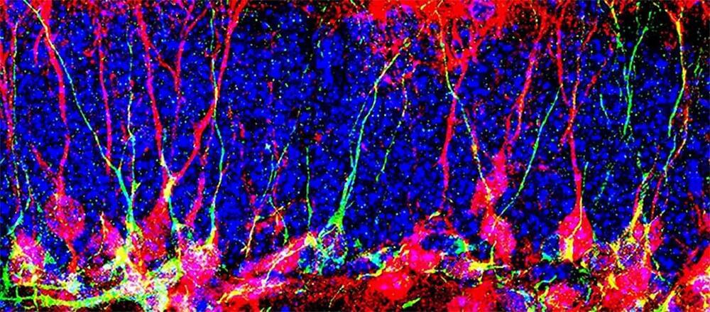
Toggle light / dark theme

Visual response shows promise as biomarker in autism-linked condition

Because the brain responses in children with different forms of autism overlapped, future therapies that are effective for Phelan-McDermid syndrome could potentially help other autistic children with similar neural patterns, Siper says.


Brain responses to visual stimuli are smaller and weaker in children with Phelan-McDermid syndrome, an autism-linked genetic condition, than in non-autistic children, according to a new study. The difference in response is greater in children with larger genetic mutations.

Mutations or deletions in SHANK3, one of the genes most strongly linked to autism, cause Phelan-McDermid syndrome. More than 80 percent of people with the condition have autism; they also often have intellectual disability, developmental delays and other medical issues, though these traits and their severity can vary widely.

The new study is the first to use electroencephalography (EEG) to measure visual evoked potentials — brain responses that occur shortly after a person views a visual stimulus — in people with Phelan-McDermid syndrome. The team previously identified differences in these responses in people with ‘idiopathic’ autism, or autism with no known genetic cause. Other researchers have linked atypical visual evoked potentials to other single-gene causes of autism, such as Rett syndrome.

This is how your brain makes your mind

What is your mind? It’s a strange question, perhaps, but if pressed, you might describe it as the part of yourself that makes you who you are—your consciousness, dreams, emotions, and memories. Scientists believed for a long time that such aspects of the mind had specific brain locations, like a circuit for fear, a region for memory, and so on.

But in recent years we’ve learned that the human brain is actually a master of deception, and your experiences and actions do not reveal its inner workings. Your mind is in fact an ongoing construction of your brain, your body, and the surrounding world.

Estimates of Americans with long COVID-19, per state

About 11.1 million Americans are living with long COVID-19, according to new estimates from The American Academy of Physical Medicine and Rehabilitation.

Long COVID-19, or persistent symptoms up to six months after being cleared of the illness, affects around 30 percent of individuals who had COVID-19, according to two recent publications from the Journal of the American Medical Association. Symptoms of long COVID-19 are varied and may include neurological challenges, cognitive problems, shortness of breath, fatigue, pain and mobility issues.

The AAPM&R has developed a dashboard estimating long COVID-19 infections. The model assumes that 30 percent of people who recover from acute COVID-19 develop long COVID-19, but users can adjust estimates based on higher or lower percentages. U.S. case data is pulled from Baltimore-based Johns Hopkins University COVID-19 data. U.S. census data uses2019estimates.

New method greatly improves X-ray nanotomography resolution

It’s been a truth for a long time: if you want to study the movement and behavior of single atoms, electron microscopy can give you what X-rays can’t. X-rays are good at penetrating into samples—they allow you to see what happens inside batteries as they charge and discharge, for example—but historically they have not been able to spatially image with the same precision electrons can.

But scientists are working to improve the image resolution of X-ray techniques. One such method is X-ray tomography, which enables non-invasive imaging of the inside of materials. If you want to map the intricacies of a microcircuit, for example, or trace the neurons in a brain without destroying the material you are looking at, you need X-ray tomography, and the better the resolution, the smaller the phenomena you can trace with the X-ray beam.

To that end, a group of scientists led by the U.S. Department of Energy’s (DOE) Argonne National Laboratory has created a new method for improving the resolution of hard X-ray nanotomography. (Nanotomography is X-ray imaging on the scale of nanometers. For comparison, an average human hair is 100,000 nanometers wide.) The team constructed a high-resolution X-ray microscope using the powerful X-ray beams of the Advanced Photon Source (APS) and created new computer algorithms to compensate for issues encountered at tiny scales. Using this method, the team achieved a resolution below 10 nanometers.

Genes for Alcohol Use Disorder and Alzheimer’s Risk Overlap: Study

ABOVE: MAPT, one of the genes linked to both heavy drinking and neurodegenerative diseases, codes for the protein tau (blue in this illustration) inside a neuron. NATIONAL INSTITUTE ON AGING/ NATIONAL INSTITUTES OF HEALTH

Some genetic risk factors for alcohol use disorder overlap with those for neurodegenerative diseases like Alzheimer’s, scientists reported in Nature Communications on August 20. The study, which relied on a combination of genetic, transcriptomic, and epigenetic data, also offers insight into the molecular commonalities among these disorders, and their connections to immune disfunction.

“By meshing findings from genome wide association studies… ith gene expression in brain and other tissues, this new study has prioritized genes likely to harbor regulatory variants influencing risk of Alcohol Use Disorder,” writes David Goldman, a neurogenetics researcher at the National Institute on Alcohol Abuse and Alcoholism (NIAAA), in an email to The Scientist. “Several of these genes are also associated with neurodegenerative disorders—an intriguing connection because of alcohol’s ability to prematurely age the brain.”

Mental Phenomena Don’t Map Into the Brain as Expected

Neuroscientists are the cartographers of the brain’s diverse domains and territories — the features and activities that define them, the roads and highways that connect them, and the boundaries that delineate them. Toward the front of the brain, just behind the forehead, is the prefrontal cortex, celebrated as the seat of judgment. Behind it lies the motor cortex, responsible for planning and coordinating movement. To the sides: the temporal lobes, crucial for memory and the processing of emotion. Above them, the somatosensory cortex; behind them, the visual cortex.

Not only do researchers often depict the brain and its functions much as mapmakers might draw nations on continents, but they do so “the way old-fashioned mapmakers” did, according to Lisa Feldman Barrett, a psychologist at Northeastern University. “They parse the brain in terms of what they’re interested in psychologically or mentally or behaviorally,” and then they assign the functions to different networks of neurons “as if they’re Lego blocks, as if there are firm boundaries there.”

But a brain map with neat borders is not just oversimplified — it’s misleading. “Scientists for over 100 years have searched fruitlessly for brain boundaries between thinking, feeling, deciding, remembering, moving and other everyday experiences,” Barrett said. A host of recent neurological studies further confirm that these mental categories “are poor guides for understanding how brains are structured or how they work.”

Prozac Changes Fat Composition of the Brain

Lipids are abundant in the brain, where they are found not just in the cell membranes of neurons, whose properties they modulate, but also in the so-called myelin sheaths insulating axons — the brain’s ‘wiring.’ The brain is therefore a surprisingly ‘fat’ organ — in fact, it is nearly 60% fat, the study’s first author, Anna Tkachev from Skoltech, said.


Summary: Prozac reduced polyunsaturated fatty acid lipid concentrations in the brains of juvenile macaque monkeys.

Source: Skoltech

Skoltech researchers and their colleagues from Russia, Germany, and the U.S. have found Prozac to reduce lipid concentrations in juvenile macaques who received the antidepressant for two years, compared to a control group of untreated animals.

While none of the monkeys in the study were depressed, the findings still offer a plausible biochemical explanation for the drug’s side effects, particularly in young patients. The paper was published in the International Journal of Molecular Sciences.

/* */