Toggle light / dark theme

Scientists at Rutgers University-Newark have discovered that when a key protein needed to generate new brain cells during prenatal and early childhood development is missing, part of the brain goes haywire—causing an imbalance in its circuitry that can lead to long-term cognitive and movement behaviors characteristic of autism spectrum disorder.

“During , there is a coordinated series of events that have to occur at the right time and the right place in order to establish the appropriate number of cells with the right connections,” said Juan Pablo Zanin, Rutgers-Newark research associate and lead author on a paper published in the Journal of Neuroscience.” Each of these steps is carefully regulated and if any of these steps are not regulated correctly, this can impact behavior.”

Zanin has been working with Wilma Friedman, professor of cellular neurobiology in the Department of Biological Sciences, studying the p75NTR —needed to regulate —to determine its exact function in brain development, gain a better understanding of how this genetic mutation could cause to die off and discover whether there is a genetic link to autism or like Alzheimer’s.

Johns Hopkins researchers report that a type of biodegradable, lab-engineered nanoparticle they fashioned can successfully deliver a “suicide gene” to pediatric brain tumor cells implanted in the brains of mice. The poly(beta-amino ester) nanoparticles, known as PBAEs, were part of a treatment that also used a drug to kill the cells and prolong the test animals’ survival.

In their study, described in a report published January 2020 in the journal Nanomedicine: Nanotechnology, Biology and Medicine, the researchers caution that for safety and biological reasons, it is unlikely that the herpes simplex virus type I thymidine kinase (HSVtk)—which makes tumor cells more sensitive to the lethal effects of the anti-viral drug ganciclovir—could be the exact therapy used to treat human medulloblastoma and atypical teratoid/rhabdoid tumors (AT/RT) in children.

So-called “suicide ” have been studied and used in cancer treatments for more than 25 years. The HSVtk gene makes an enzyme that helps restore the function of natural tumor suppression.

A new unique signal discovered within the brain might be what makes us human:

https://science.sciencemag.org/content/367/6473/83

For the latest news in neuroscience, psychology, and artificial intelligence, please like and follow our Facebook page: https://www.facebook.com/The-Neuro-Network-383136302314720/


A special developmental program in the human brain drives the disproportionate thickening of cortical layer 2/3. This suggests that the expansion of layer 2/3, along with its numerous neurons and their large dendrites, may contribute to what makes us human. Gidon et al. thus investigated the dendritic physiology of layer 2/3 pyramidal neurons in slices taken from surgically resected brain tissue in epilepsy patients. Dual somatodendritic recordings revealed previously unknown classes of action potentials in the dendrites of these neurons, which make their activity far more complex than has been previously thought. These action potentials allow single neurons to solve two long-standing computational problems in neuroscience that were considered to require multilayer neural networks.

Science, this issue p. 83

The active electrical properties of dendrites shape neuronal input and output and are fundamental to brain function. However, our knowledge of active dendrites has been almost entirely acquired from studies of rodents. In this work, we investigated the dendrites of layer 2 and 3 (L2/3) pyramidal neurons of the human cerebral cortex ex vivo. In these neurons, we discovered a class of calcium-mediated dendritic action potentials (dCaAPs) whose waveform and effects on neuronal output have not been previously described. In contrast to typical all-or-none action potentials, dCaAPs were graded; their amplitudes were maximal for threshold-level stimuli but dampened for stronger stimuli. These dCaAPs enabled the dendrites of individual human neocortical pyramidal neurons to classify linearly nonseparable inputs—a computation conventionally thought to require multilayered networks.

The size of the human brain increased profoundly during evolution. A certain gene that is only found in humans triggers brain stem cells to form a larger pool of stem cells. As a consequence, more neurons can arise, which paves the way to a bigger brain. This brain size gene is called ARHGAP11B and so far, how it works was completely unknown. Researchers at the Max Planck Institute of Molecular Cell Biology and Genetics in Dresden now uncovered its mode of action. They show that the ARHGAP11B protein is located in the powerhouse of the cell—the mitochondria—and induces a metabolic pathway in the brain stem cells that is characteristic of cancer cells.

The research group of Wieland Huttner, a founding director of the Max Planck Institute of Molecular Cell Biology and Genetics, has been investigating the underlying the expansion of the brain during mammalian evolution for many years. In 2015, the group reported a key role for a gene that is only present in humans and in our closest extinct relatives, the Neanderthals and Denisovans. This gene, named ARHGAP11B, causes the so-called basal brain stem to expand in number and to eventually increase the production of neurons, leading to a bigger and more folded brain in the end. How the gene functions within the basal brain stem cells has been unknown so far.

Takashi Namba, a postdoctoral scientist in the research group of Wieland Huttner, wanted to find the answer to this question, together with colleagues from the Max Planck Institute, the University Hospital Carl Gustav Carus Dresden, and the Department of Medical Biochemistry at the Semmelweis University, Budapest. He found that the ARHGAP11B protein is located in mitochondria, the organelles that generate most of the cell’s source of chemical energy and hence are often referred to as the powerhouse of the cell. Takashi Namba explains the results: We found that ARHGAP11B interacts with a protein in the membrane of mitochondria that regulates a membrane pore. As a consequence of this interaction, the pores in the membrane are closing up, preventing calcium leakage from the mitochondria. The resulting higher calcium concentration causes the mitochondria to generate chemical energy by a metabolic pathway called glutaminolysis.

In the story of Goldilocks, a little girl tastes three different bowls of porridge to find which is not too hot, not too cold, but just the right temperature. In a study published in Advanced Therapeutics, University of Minnesota Medical School researchers report on a “Goldilocks” balance which holds the key to awakening the body’s immune response to fight off brain cancer.

The most common form of adult is glioblastoma. Doctors diagnose about 14,000 glioblastoma cases in the U.S. each year. This aggressive cancer has claimed the lives of Senators John McCain and Edward Kennedy.

“Our body has armies of white blood cells that help us fight off bacteria, viruses and cancer cells. This constellation of cells constitute our immune system,” said senior author Clark C. Chen, MD, Ph.D., Lyle French Chair in Neurosurgery and Head of the Department of Neurosurgery at the University of Minnesota Medical School. “One of the key reasons why glioblastoma is so aggressive is that it shuts off this immune system.”

Alzheimer’s disease could be reversed by shining light directly into the brain through the nose and skull, scientists believe.

The first major trial to see if light therapy could be beneficial for dementia has just begun following astonishing early results which have seen people regain their memory, reading and writing skills, and orientation.

If successful it would be the first treatment to actually reverse the disease. So far, even the most hopeful drugs, such as Biogen’s aducanumab, have only managed to slow the onset of dementia, and many scientists had given up hope of reversing brain damage once it had already happened.

Specifically, AstroRx is a cell therapy product containing functional healthy astrocytes derived from human embryonic stem cells that aim to protect diseased motor neurons. The cells are injected in the patient through the spinal canal.

The company initiated its first ALS clinical trials in March 2018.

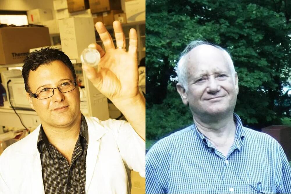
Prof. Michel Revel, founder and CEO of the company and winner of both the EMET Prize and the Israel Prize, said that the clinical trial results “provide the confidence needed to move forward” with cohorts B and C, which he said both “hold the potential for a prolonged response.”