Toggle light / dark theme

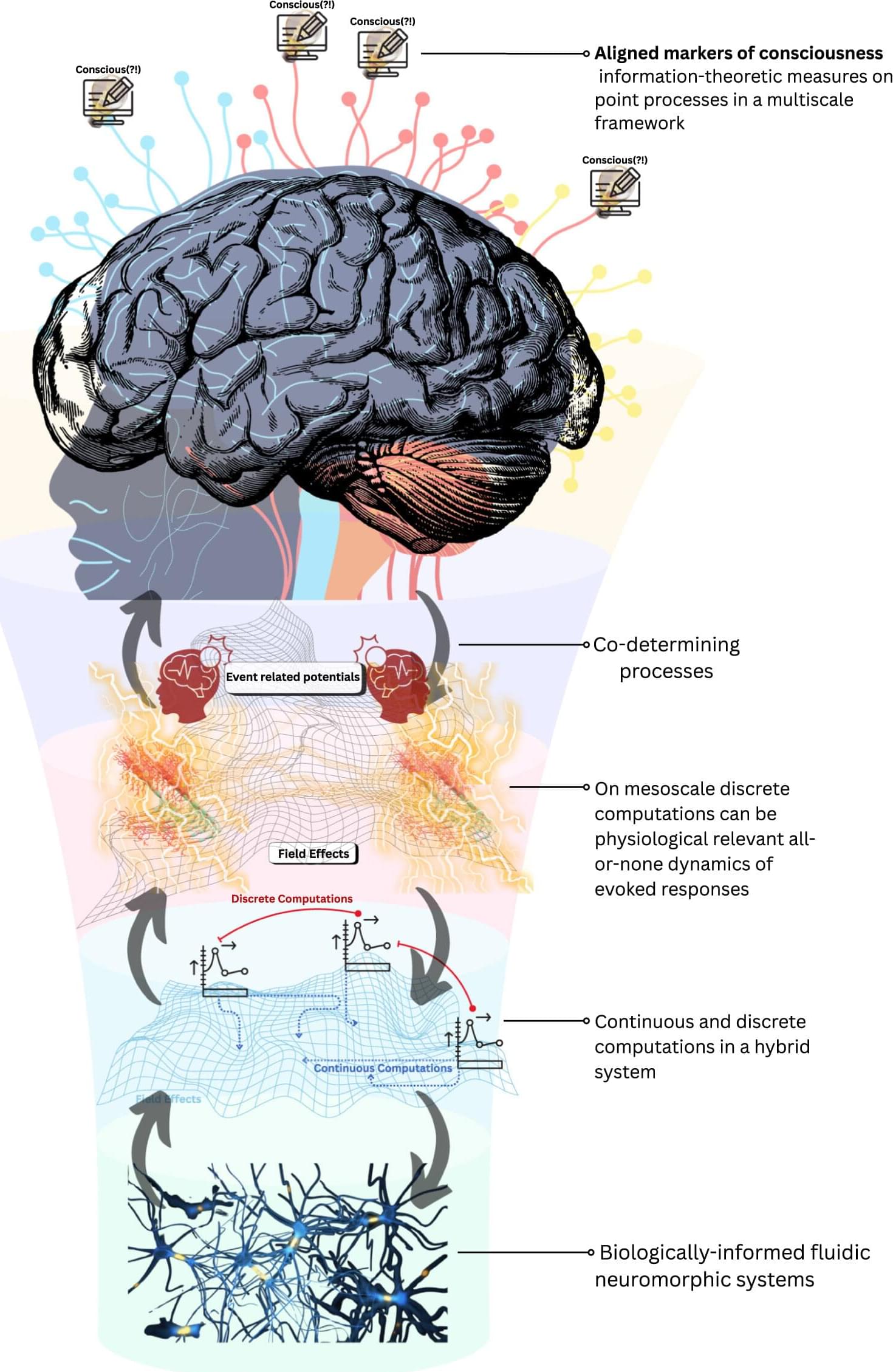
Our Conscious Perception of the World Depends on This Deep Brain Structure

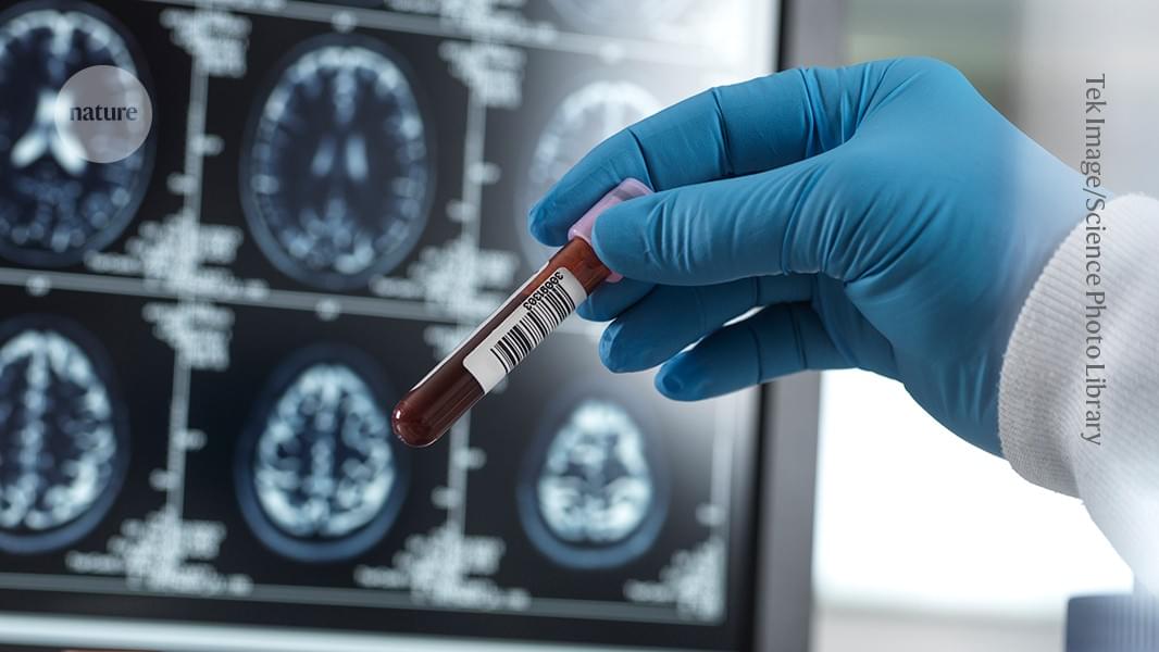
The study is “really pretty remarkable,” said Christopher Whyte at the University of Sydney, who was not involved in the work, to Nature. One of the first to simultaneously record activity in both deep and surface brain regions in humans, it reveals how signals travel across the brain to support consciousness.

Consciousness has teased the minds of philosophers and scientists for centuries. Thanks to modern brain mapping technologies, researchers are beginning to hunt down its neural underpinnings.

At least half a dozen theories now exist, two of which are going head-to-head in a global research effort using standardized tests to probe how awareness emerges in the human brain. The results, alongside other work, could potentially build a unified theory of consciousness.

Brain networks study examines the transition from early to chronic psychosis

Patients in the early stages of psychosis respond to treatments differently than those who have developed a chronic version of the disorder. Understanding the neurobiological changes from early to chronic stages is essential for developing targeted prevention and treatment strategies. But how symptoms change during this transition—and what role the brain plays—is unclear.

Researchers at Yale School of Medicine (YSM) have now examined patients with early and chronic forms of psychosis to map symptom evolution and identify relevant brain networks. They published their findings in the journal Neuropsychopharmacology.

“We are interested in how psychosis and psychiatric disorders develop,” says Maya Foster, first author of the study and a Ph.D. student in the lab of Dustin Scheinost, Ph.D., associate professor of radiology and biomedical imaging at YSM.

Psychosis-inducing lesions affect a common brain circuit in the hippocampus, research reveals

A new study investigated the brain circuits involved in psychosis—a condition characterized by delusions, hallucinations, disorganized thinking and detachment from reality.

Andrew Pines, MD, MA, a resident in the Department of Psychiatry at Brigham and Women’s Hospital and a researcher in the Center for Brain Circuit Therapeutics, is the lead author of the paper published in JAMA Psychiatry titled “Mapping Lesions That Cause Psychosis to a Human Brain Circuit and Proposed Stimulation Target.”

Psychosis is the classic symptom of schizophrenia, a that causes marked disability in otherwise young and healthy patients. The researchers analyzed published cases in which focal brain damage caused psychosis, with the idea that if damaging a brain circuit causes a symptom, then mapping that circuit might tell us about how to treat that symptom.

Emergence of psychosis in Alzheimer disease links to elevations in p-tau181

For individuals with Alzheimer disease (AD), the emergence of psychosis is associated with elevations in levels of plasma tau phosphorylated at threonine 181 (p-tau181), according to a study published online June 26 in JAMA Psychiatry.

Jesus J. Gomar, Ph.D., and Jeremy Koppel, M.D., from the Feinstein Institutes for Medical Research in Manhasset, New York, examined the longitudinal dynamics of p-tau181 and neurofilament light chain protein (NfL) levels in association with the emergence of psychotic symptoms. Patients with (MCI) and AD (with psychosis [AD+P] and without psychosis [AD−P]) and participants who were cognitively unimpaired (CU) were compared at baseline.

For the longitudinal analysis, participants with MCI and AD were categorized into those with evidence of psychosis at baseline and those who showed incidence of psychosis over the course of the study. The cohort included 752 participants with AD and 424 CU participants.

In Down syndrome mice, 40Hz light and sound improve cognition, neurogenesis and connectivity

Studies by a growing number of labs have identified neurological health benefits from exposing human volunteers or animal models to light, sound and/or tactile stimulation at the brain’s “gamma” frequency rhythm of 40Hz. In the latest such research at The Picower Institute for Learning and Memory and Alana Down Syndrome Center at MIT, scientists found that 40Hz sensory stimulation improved cognition and circuit connectivity and encouraged the growth of new neurons in mice genetically engineered to model Down syndrome.

Li-Huei Tsai, Picower Professor at MIT and senior author of the new study in PLOS ONE, said that the results are encouraging but also cautioned that much more work is needed to test whether the method, called GENUS (for Gamma Entrainment Using Sensory Stimulation), could provide clinical benefits for people with Down syndrome. Her lab has begun a small study with human volunteers at MIT.

“While this work, for the first time, shows the beneficial effects of GENUS on Down syndrome using an imperfect mouse model, we need to be cautious as there is not yet data showing whether this also works in humans,” said Tsai, who directs The Picower Institute and The Alana Center, and is a member of MIT’s Brain and Cognitive Sciences faculty.

The gene responsible for cognitive defects in Down syndrome

Learning and memory impairments in a Down syndrome mouse model were reversed by correcting expression of a gene that influences the generation of new neurons in the brain. The finding could pave the way to treat the cognitive impairment associated with the syndrome in humans.

Adult neurogenesis is the process of generating new neurons in the adult brain. Defects in this process have been observed in various animal models of neurological disorders including schizophrenia, depression, Parkinson’s disease, Alzheimer’s disease, and such as Down syndrome. But the precise cellular and underlying adult neurogenesis and their links to neurological disorders are not well understood.

Molecular neurobiologist Kyung-Tai Min at Korea’s Ulsan National Institute of Science and Technology and his colleagues found that interactions between a gene called the Down syndrome critical region 1 (DSCR1) and two other molecules, TET1 and miRNA-124, were necessary for adult neurogenesis and were important in learning and memory.

Death is not final, it can be reversed claims New York University doctor

Sam Parnia, associate professor at New York University, suggests that death can be reversed and our brains may remain salvageable for hours or days after death. He emphasizes that death can be viewed as an injury process, with the potential for revival through ECMO machines and specific drugs used in CPR cocktails to aid recovery.

Scientists create first reprogrammed brain cells that could cure Alzheimer’s

In a world-first, scientists have figured out how to reprogram cells to fight — and potentially reverse — brain diseases like Alzheimer’s.

Researchers at the University of California, Irvine created lab-grown immune cells that can track down toxic brain buildup and clear it away, restoring memory and brain function in mice.

They did this by turning stem cells — which can become any cell in the body — into brain immune cells called microglia.