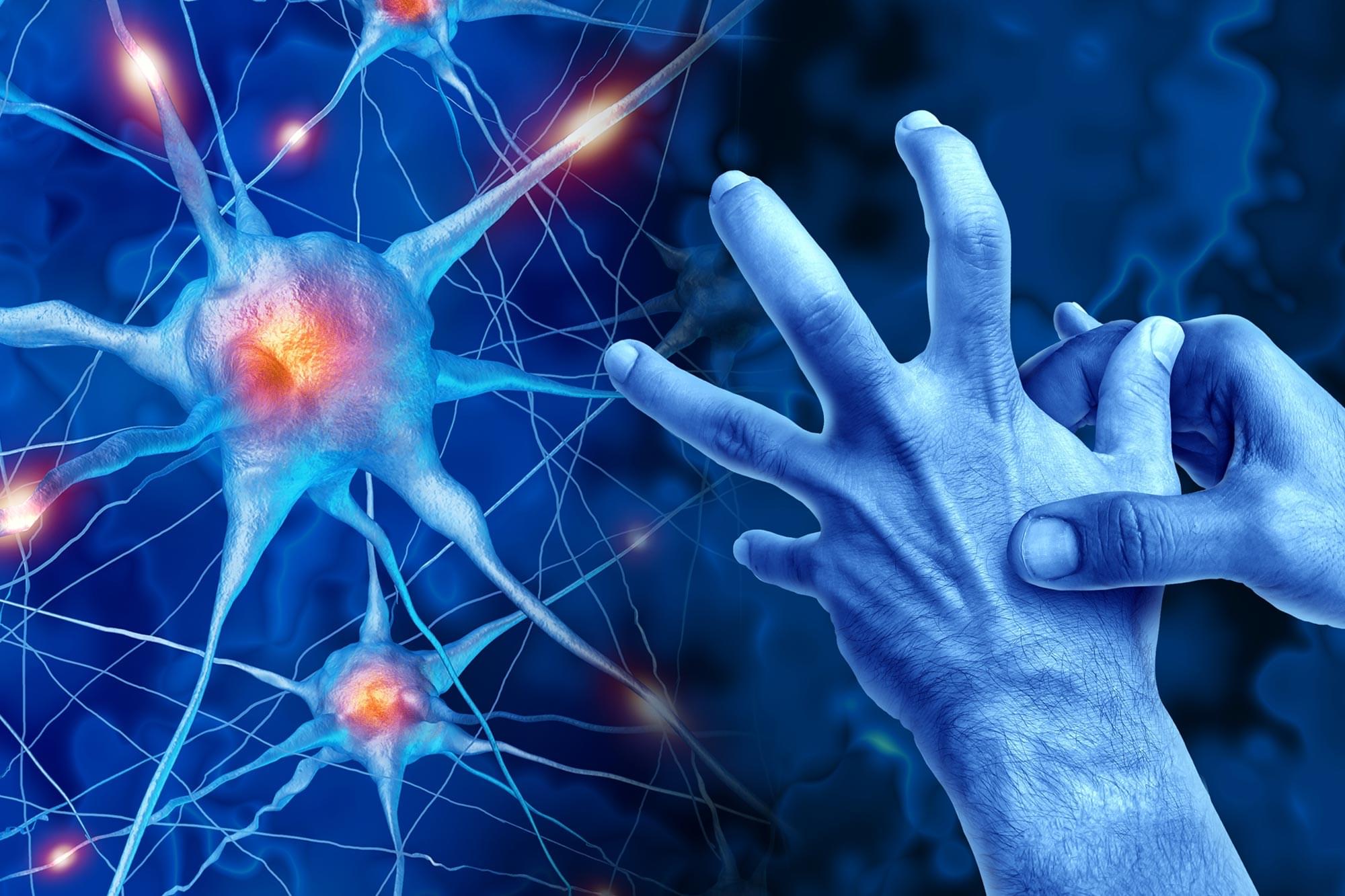
Researchers have identified two proteins on the surface of neurons that may help drive the spread of Parkinson’s disease in the brain.
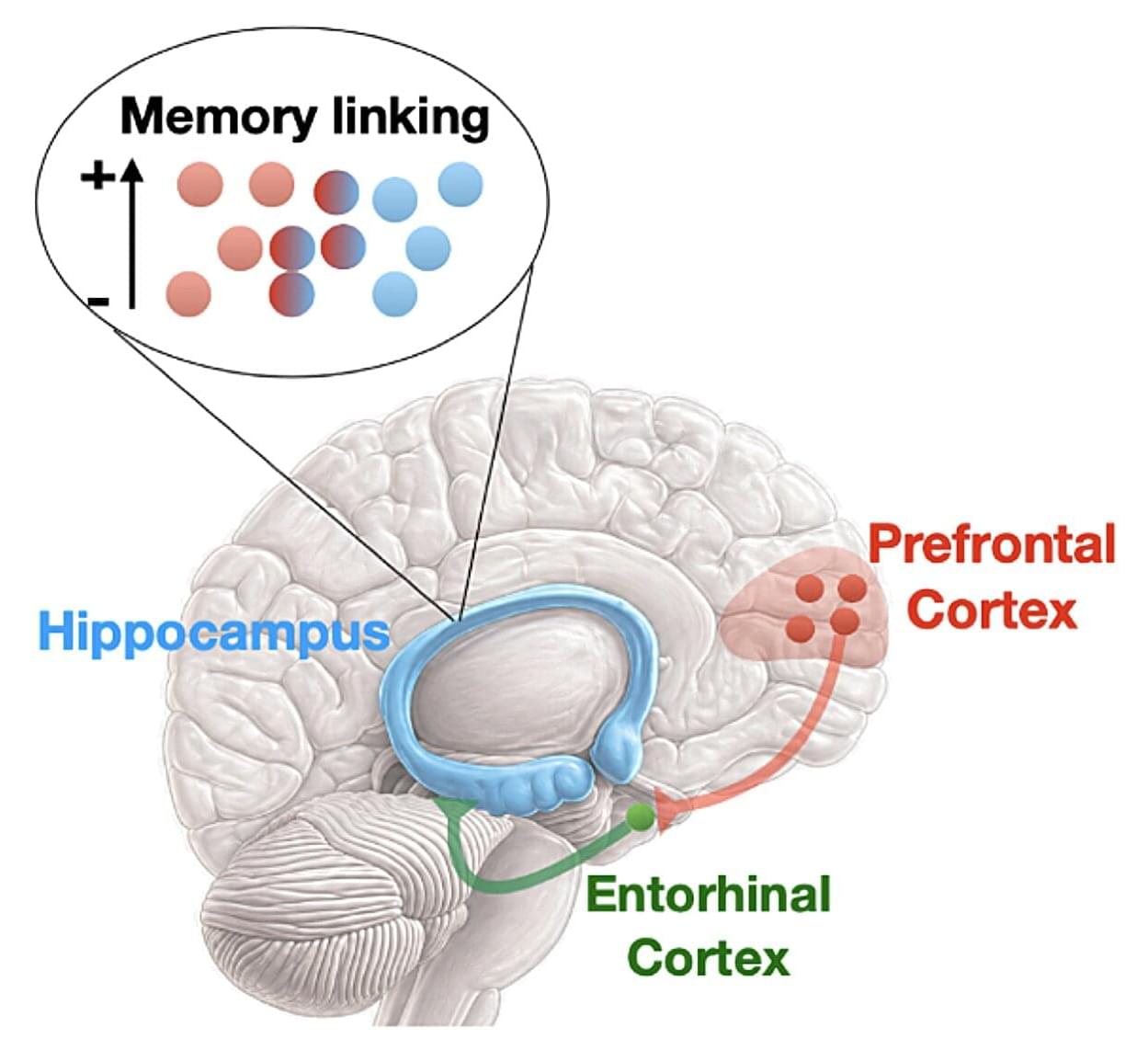
Our memories of past events are typically not isolated, but they are linked to other related memories. This ability to establish connections between related memories is highly advantageous, as it helps us to recognize familiar patterns in new situations and make predictions that can inform our decisions.
Researchers at UCLA’s Brain Research Institute recently carried out a study on mice aimed at better understanding how the brain decides what memories are connected and which ones are not. Their paper, published in Nature Neuroscience, pinpoints brain regions that could play a role in the organization of memories into coherent pools of knowledge.
“Our lab has long been interested in understanding how the brain connects related memories,” André F. de Sousa, first author of the paper, told Medical Xpress. “In everyday life, new experiences are rarely processed in isolation. Instead, they are often shaped by what we have learned before. This ability allows us to link related events, build knowledge, and use past experiences to guide future behavior. However, this process needs to be carefully controlled.”
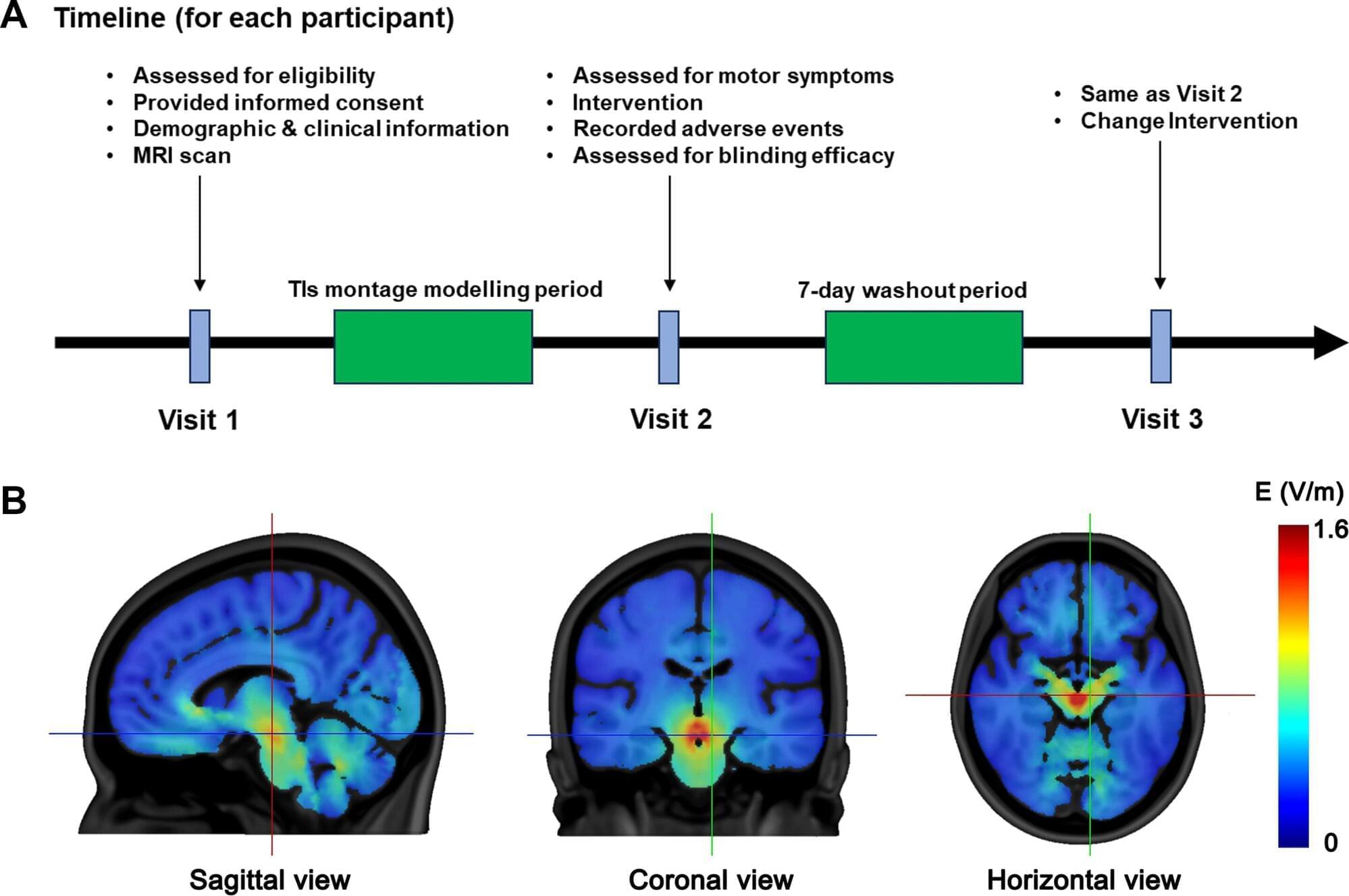
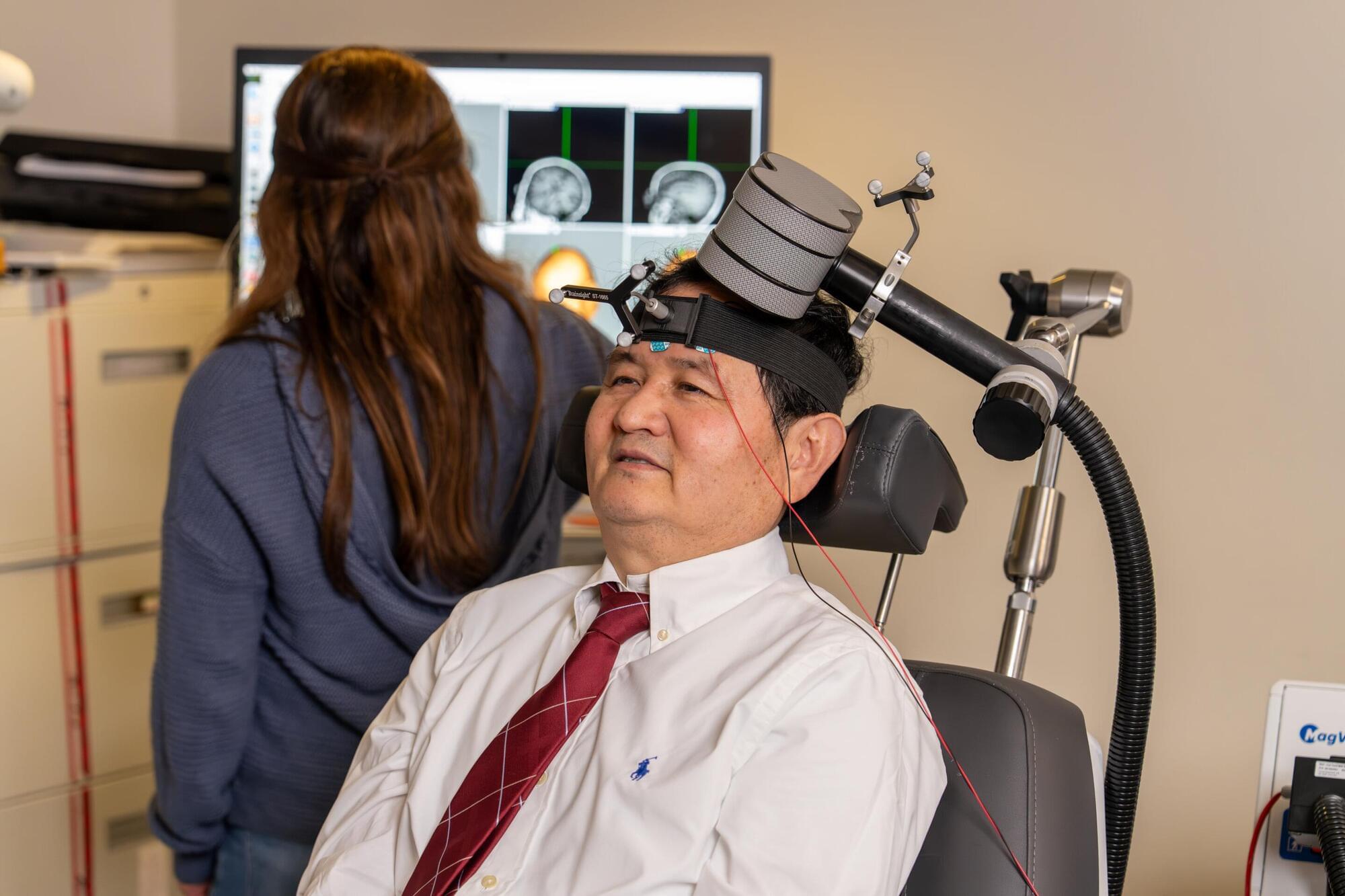
A novel, noninvasive brain stimulation approach—known as transcranial temporal interference stimulation (TIs)—may offer a new way to treat motor symptoms in Parkinson’s disease without the need for surgery, according to a pilot study appearing in eBioMedicine. The technique, which uses overlapping electrical currents to selectively target deep brain regions, significantly improved movement in patients compared with a sham treatment when targeting the subthalamic nucleus.
Parkinson’s disease is a progressive neurological disorder that affects movement, often causing tremor, stiffness, and slowed motion. One of the most effective treatments for advanced symptoms is deep brain stimulation (DBS), which involves implanting electrodes into the brain. TIs may be able to achieve a similar effect—targeting the same deep brain structures —but entirely from outside the skull, using carefully calibrated electrical fields delivered through the scalp.
In the randomized, double-blind, crossover study, titled “Transcranial temporal interference stimulation targeting the subthalamic region for motor symptoms in Parkinson’s disease: a pilot, randomised, double-blind, sham-controlled crossover study,” 30 people with early-to mid-stage Parkinson’s disease received a single 20-minute session of individualized TIs targeting the subthalamic region—a key node in the brain’s motor control network—as well as a sham or placebo treatment in a separate session.
For many people who smoke, quitting is not just a matter of willpower. It is a tug-of-war in the brain—between the pull of reward and the ability to resist.
A study published in the Journal of Psychiatric Research suggests that shifting that balance may be possible. Using a noninvasive brain stimulation technique called repetitive transcranial magnetic stimulation, or rTMS, researchers at MUSC Hollings Cancer Center found that stimulating a specific brain region that regulates self-control significantly reduced how much people smoked.
When danger lurks, instinct keeps us safe. It compels us to run from a burning building or wrestle a knife-wielding attacker to the ground. It also adjusts our body physiology to support these behaviors.
Survival helps explain why. But the mechanisms that link the brain and the body—the “switch” between rest and action—have long been shrouded in mystery.
A research team at Rutgers University-New Brunswick thinks they may have identified a key mechanism, and the findings may hold important clues to how diverse neurological conditions, such as alcohol use disorder and Parkinson’s, could be diagnosed and treated.
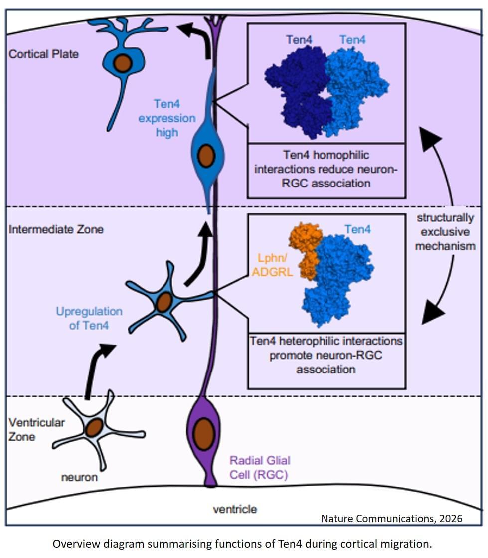
During brain development, neurons can regulate their movement until they reach their final destination thanks to a “molecular switch” involving the protein Teneurin 4 (Ten4). This protein can guide neuronal migration through mutually exclusive molecular pathways and determine the direction of nerve cells.
The discovery, published in the journal Nature Communications, improves our understanding of the molecular mechanisms that control neuronal migration and offers new insights into how the brain develops at the molecular level.
The study combines advanced techniques — structural protein studies, gene editing in animal models and super-resolution microscopy — to broaden our understanding of the origins of neurodevelopmental disorders and psychiatric or neurological conditions —schizophrenia, epilepsy, autism, bipolar disorder, etc. — which may be linked to errors in neuronal migration.
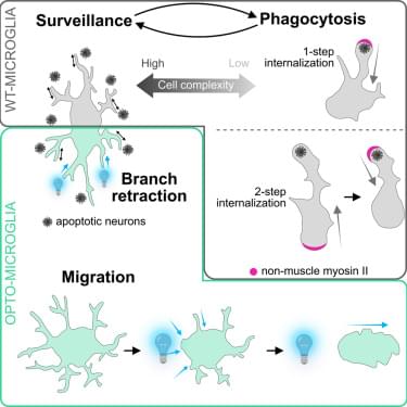
Biermeier et al. use live imaging in zebrafish to show that microglia alternate between distinct morphological states that support brain surveillance and phagocytosis. By optogenetically controlling cytoskeletal contractility, they demonstrate programmable, reversible control of microglial behavior in the living brain.

You may think you’re the protagonist of your own story. According to Oxford physicist Vlatko Vedral, however, you’re more like a puppet — whose strings are being pulled into a million parallel universes at any given time.
As Vedral argues in a recent issue of Popular Mechanics, the pop-sci version of the “observer effect” — where the act of observation or measurement affects a system — gets the cause-and-effect backward. The typical story goes something like this: quantum objects hang out in multiple states at once, until some observer glances over. At this point, the multiple states collapse and only one is left, an assumption that can lead various woo-woo interpretations, like that we create reality simply by observing it.
Physics, Verdal says, does not support that idea. That collapse effect isn’t a special power of human consciousness, but rather a fact of physics that says interactions — any interaction — forces a quantum system to commit to a definite state.