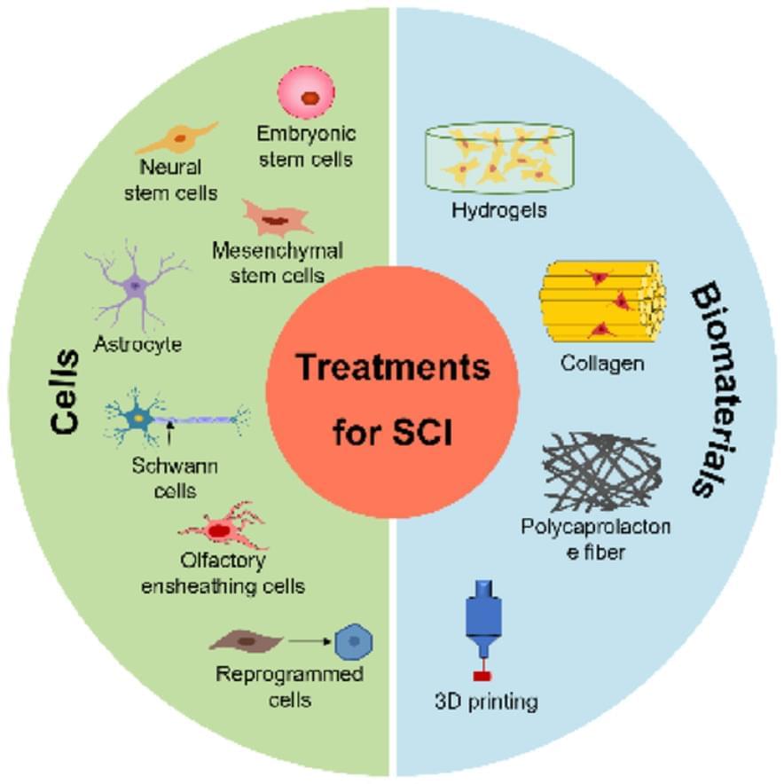
Toggle light / dark theme

Research published in Nature has revealed that neural computations in different individuals can be implemented to solve the same decision-making tasks, even when the behavioral outcomes appear identical.

Cognitive flexibility is the ability of a brain to adapt its response to the same , like light or sound, based on different contexts. For example, if someone calls your name in a crowded room, you must focus on the sound’s location or the voice characteristics to identify the person. This flexibility in selecting and processing while ignoring irrelevant information is crucial for survival and effective interaction with our environment.

While previously studied, the individual in neural computations yielding the same outcomes is poorly understood and lacks a comprehensive framework. The researchers in the Nature study aimed to understand these mechanisms.

Slow-wave sleep plays a crucial role in strengthening memory by enhancing synaptic connections in the brain, with new findings suggesting potential methods for boosting memory through targeted stimulation.

For nearly two decades, scientists have known that slow, synchronized electrical waves in the brain during deep sleep play a key role in forming memories. However, the underlying reason remained unclear — until now. In a new study published in Nature Communications, researchers from Charité – Universitätsmedizin Berlin propose an explanation. They found that these slow waves make the neocortex, the brain’s long-term memory center, especially receptive to new information. This discovery could pave the way for more effective memory-enhancing treatments in the future.

How Memories Form During Sleep

Aging is an inevitable aspect of life, but age-related diseases are not an inseparable part of the aging process, and their risk can be reduced through a healthy lifestyle. Vitamin K has a broader impact than just blood clotting, and yet it remains overshadowed by other vitamins and underestimated by both doctors and consumers. Vitamin K (VK) is a multifunctional micronutrient with anti-inflammatory and antioxidant properties, whose deficiency may cause age-related diseases such as cardiovascular diseases, neurodegenerative diseases and osteoporosis. There is a growing body of evidence supporting the role of vitamin K as a protective nutrient in aging and inflammation. This review summarizes the current knowledge regarding the molecular aspects of the protective role of vitamin K in aging and age-related diseases and its clinical implications.

Salt, or more precisely the sodium it contains, is very much a “Goldilocks” nutrient. Low sodium levels cause a drop in blood volume, which can have serious, sometimes deadly, health consequences. Conversely, too much salt can lead to high blood pressure and cardiovascular disease.

In modern America, where most people consume a , almost no one is in danger of having too little salt. However, given the critical importance of sodium for body and brain functions, evolution has developed a powerful drive to consume salt in situations where there is a deficiency.

Understanding the brain circuitry that controls salt appetite has proved elusive, but now a new study by University of Iowa researchers has identified the first and, thus far, only neurons necessary for salt appetite.

As the global population ages, more of us face frightening cognitive decline, personally or in our loved ones. There are over 10 million new cases of dementia globally each year.

But a study published this year suggests up to 13 percent of people diagnosed with dementia in the US may have a misdiagnosis and are instead left struggling with a condition that can be treated.

“Health care providers [must be] made aware of this potential overlap between dementia and hepatic encephalopathy, which is treatable,” said Virginia Commonwealth University hepatologist Jasmohan Bajaj in July.

Personalized content and ads can also include things like video recommendations, a customized YouTube homepage, and tailored ads based on past activity, like the videos you watch and the things you search for on YouTube. We also use cookies and data to tailor the experience to be age-appropriate, if relevant.

Select “More options” to see additional information, including details about managing your privacy settings. You can also visit g.co/privacytools at any time.

Considering what’s known about their brain structures, sensory systems and learning capacity, it appears that cephalopods as a group may be similar in intelligence to vertebrates as a group. Since many societies have animal welfare standards for mice, rats, chickens and other vertebrates, logic would suggest that there’s an equal case for regulations enforcing humane treatment of cephalopods.

Such rules generally specify that when a species is held in captivity, its housing conditions should support the animal’s welfare and natural behavior. This view has led some U.S. states to outlaw confined cages for egg-laying hens and crates too narrow for pregnant sows to turn around.

Animal welfare regulations say little about invertebrates, but guidelines for the care and use of captive cephalopods have started to appear over the past decade. In 2010, the European Union required considering ethical issues when using cephalopods for research. And in 2015, AAALAC International, an international accreditation organization for ethical animal research, and the Federation of European Laboratory Animal Science Associations promoted guidelines for the care and use of cephalopods in research. The U.S. National Institutes of Health is currently considering similar guidelines.

Penn Engineers have modified lipid nanoparticles (LNPs)—the revolutionary technology behind the COVID-19 mRNA vaccines—to not only cross the blood-brain barrier (BBB) but also to target specific types of cells, including neurons. This breakthrough marks a significant step toward potential next-generation treatments for neurological diseases like Alzheimer’s and Parkinson’s.

In a new paper in Nano Letters, the researchers demonstrate how —short strings of —can serve as precise targeting molecules, enabling LNPs to deliver mRNA specifically to the that line the blood vessels of the brain, as well as neurons.

This represents an important advance in delivering mRNA to the cell types that would be key in treating neurodegenerative diseases; any such treatments will need to ensure that mRNA arrives at the correct location. Previous work by the same researchers proved that LNPs can cross the BBB and deliver mRNA to the brain, but did not attempt to control which cells the LNPs targeted.

In their Review article earlier this year, Fedorenko, Ivanova & Regev (Fedorenko, E., Ivanova, A. A. & Regev, T. I. The language network as a natural kind within the broader landscape of the human brain. Nat. Rev. Neurosci. 25, 289–312 (2024))1 propose a functional separation between the core language network and other perceptual, motor and higher-level cognitive components of communication-related networks in the left hemisphere of the human brain. In the ‘Open questions and a way forward’1 section that ends their Review, the authors discuss the need for cross-species comparative research to disentangle how these brain networks came to support human language. Here, we suggest that the authors’ functional separation of a core language network and other components in the human brain is grounded in the evolution of two separate structural networks within primate brains.

Fedorenko and colleagues describe the core language network as left-lateralized, and involving the middle frontal gyrus (MFG), inferior frontal gyrus (IFG), superior temporal gyrus (STG) and middle temporal gyrus (MTG). Perceptual and motor systems for speech are defined as separate subsystems located in auditory cortex and speech perception areas in the STG and motor cortex and motor planning areas1, respectively. Importantly, these functionally defined key brain areas are known to be structurally connected via dorsally and ventrally located white-matter fibre tracts, which guarantee the information flow between areas. In humans, two separate dorsal pathways that provide structural connections have been identified for two distinct networks2,3 (Fig. 1).