Watch more videos on this topic here: https://shorturl.at/eYLvNWhat is the relationship between consciousness and the nature of ultimate reality? Some say th…
Category: neuroscience – Page 56
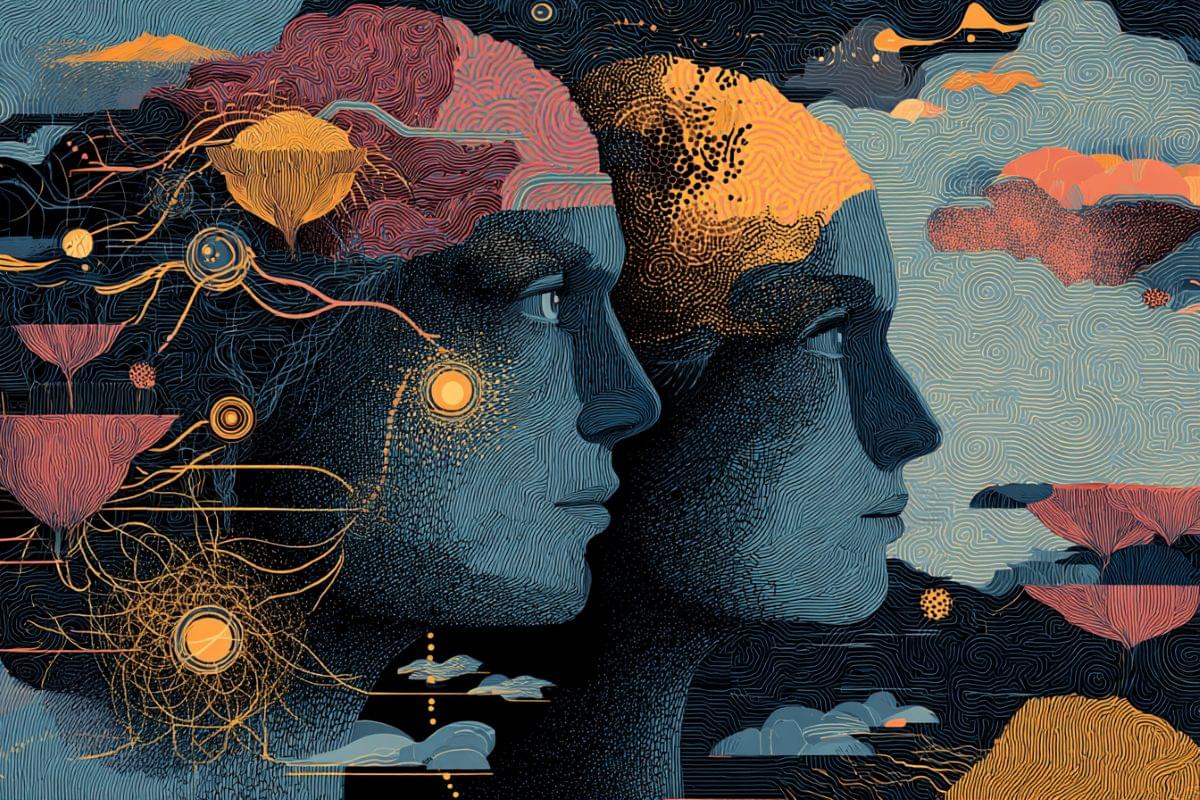
What Higher Dimensional Beings Look Like (And They’re Watching!) Per Donald Hoffman
Let’s unravel the reality beyond our space-time ♾️🔍 Go to https://piavpn.com/beeyondideas to get 83% off from our sponsor Private Internet Access with 4 months free!
Watch Part 2 of this series: • Higher-Dimensional Beings Are Staring at U…
Want to support our production? Feel free to join our membership at https://youtube.com/watch?v=FIBC5w9a5kU&si=Qy-HR0sj8USp6scG
Special thanks to our beloved YouTube members this month: Powlin Manuel, Saïd Kadi, Nate Lachae, Alison Rewell, Thomas Lapins, Ahmad Salahudin, Antonio Ferriol Colombram, Anton Nicolas Burger 🚀🚀🚀
Experts featured in this video include Donald Hoffman, Annaka Harris and Leonard Susskind.
Chapters:
The Ant and the Absolute: How Feynman Discovered Bio-Computing in a Sink
Why does an ant, with a brain smaller than a grain of sand, find the shortest path better than a human engineer?
Richard Feynman didn’t learn about ants from a textbook. He learned by sitting on his bathroom floor with a sugar cube and a stopwatch. What he discovered wasn’t just biology—it was a biological supercomputer solving the \.
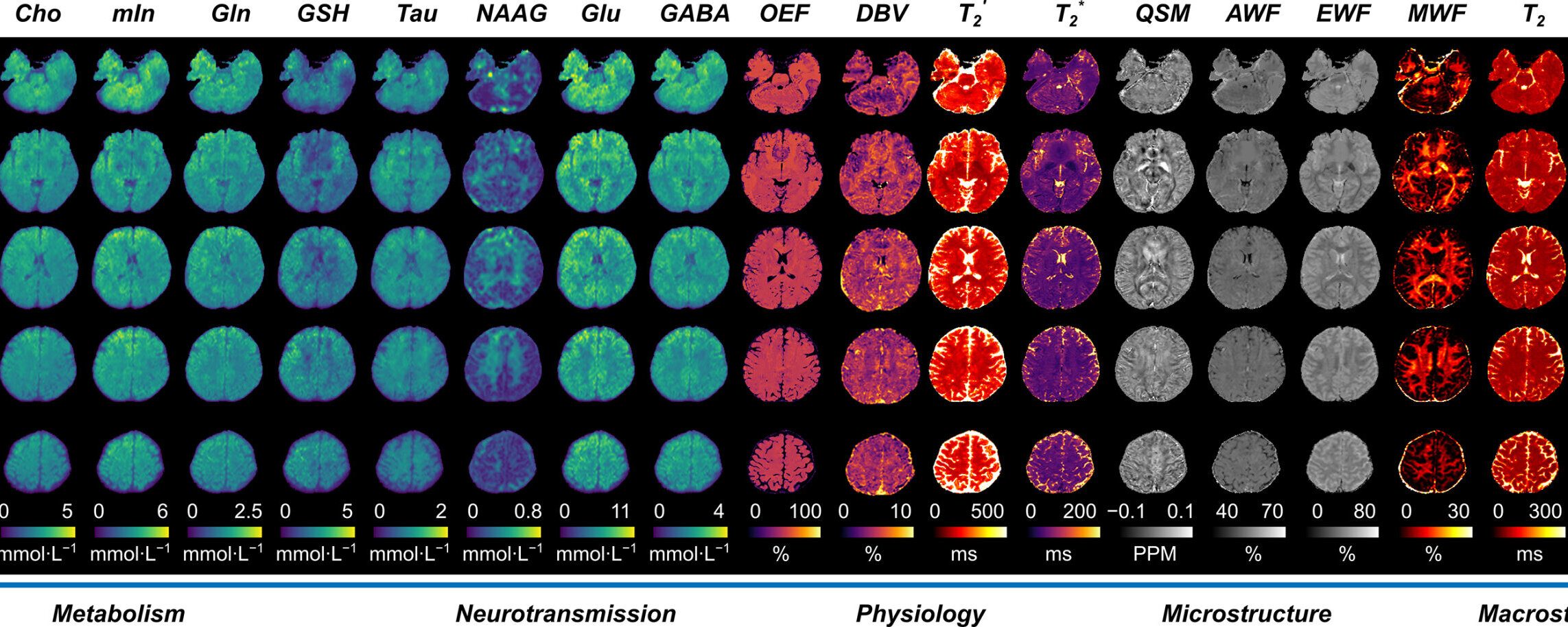
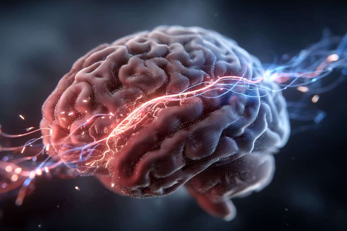
New MRI technology maps 20-plus brain biomarkers in a single 14-minute scan
New multiplexed imaging technology using standard clinical MRI systems can simultaneously map more than 20 biomarkers in high resolution, providing a comprehensive view of the brain with a single scan.
Researchers at the University of Illinois Urbana-Champaign demonstrated the multiplexed MRI technology (MRx) by characterizing brain tumors and multiple sclerosis lesions—revealing different structural, physiological and molecular changes within the diseases. Led by Zhi-Pei Liang, a professor of electrical and computer engineering and a member of the Beckman Institute for Advanced Science and Technology at the U. of I., the team has reported its findings in the journal Nature.
“MRx can be a powerful tool for noninvasive tissue characterization, helping to advance personalized, precision and predictive medicine,” Liang said. “By providing rich, multidimensional biomarkers to capture disease progression and treatment response, this capability could open new opportunities for more precise diagnosis, individualized treatment planning and improved patient outcomes.”
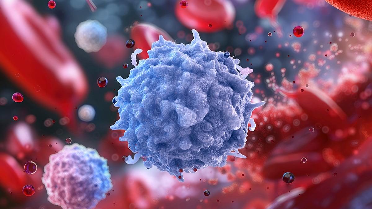
Study of a Million Blood Cells Helps Explain Why Women Face More Autoimmune Disease
Autoimmune diseases, where the body’s own immune system mistakenly goes on the attack, are much more common in women – and a new study analyzing more than 1.25 million blood cells goes a long way to explaining why.
The analysis, led by a team from the Garvan Institute of Medical Research in Australia, revealed over 1,000 genetic ‘switches’ in immune cells that work differently depending on sex.
In short, these variations in gene activity mean that inflammatory pathways that respond to threats are likely to be busier in women, leading to a greater risk of conditions like lupus and multiple sclerosis.

Digital therapy outperforms referrals to campus clinics among college students
College students with anxiety, depression and eating disorders may be more likely to start and to respond more positively to therapy offered via a digital app compared to referrals to in-person campus clinics, according to a study led by Penn State researchers and published in the journal Nature Human Behaviour.
Globally, an estimated 40% to 60% of college students experience a mental health disorder at some point, and the need for campus counseling services has increased faster than institutions’ capacity to provide these services, according to the researchers.
The research team wanted to see if a proactive intervention using a digital therapy app could effectively treat anxiety disorders, depression and eating disorders, as well as address the increased need for psychological services.