Toggle light / dark theme

Blood test can predict Alzheimer’s disease progression years before symptoms or brain scan changes

A study by investigators at Mass General Brigham has found that a blood test of plasma phosphorylated tau 217 (pTau217), an Alzheimer’s disease biomarker, can predict the progression of amyloid PET scan changes and cognitive decline in cognitively healthy older adults. The findings may help push back the clock to enable simpler, earlier disease prediction and indicate who may be at risk for cognitive decline. The results are published in Nature Communications.

“We used to think that PET scan detection was the earliest sign of Alzheimer’s disease progression, revealing amyloid accumulation in the brain 10 to 20 years before symptoms appear,” said lead author Hyun-Sik Yang, MD, a neurologist with Mass General Brigham Neuroscience Institute and an associate member of the Broad Institute of MIT and Harvard. “But now we are seeing that pTau217 can be detected years earlier, well before clear abnormalities appear on amyloid PET scans.”

Last year, the U.S. Food and Drug Administration cleared the first blood test for Alzheimer’s disease, paving the way for a cheaper, less invasive alternative to lumbar punctures and PET scans. The new study by Yang and colleagues adds important evidence about the predictive potential of these kinds of blood tests.

Proteomic insights into troponin elevation following COVID-19 infection

Background Raised cardiac troponin-I is a common finding in patients hospitalised with acute viral infections, including but not limited to COVID-19. This often occurs in the absence of overt myocardial injury presenting a challenge for interpretation. The mechanisms underlying troponin elevation are uncertain.

Methods The CISCO-19 (Cardiovascular Imaging in SARS-CoV-19) study (NCT04403607) is a prospective, multicentre cohort study, in which hospitalised PCR-confirmed COVID-19 participants (N=267) underwent multisystem evaluation at enrolment and at 28–60 days. The study incorporated plasma proteomics (SOMAscan V.4.1), cardiovascular MRI and clinical biomarkers. Of these, 211 had baseline plasma proteomic data and 185 completed follow-up sampling. Matched proteomic and imaging data were available for 155 participants (mean age: 55 years (SD 12); 43% female).

Results A high likelihood of myocarditis was identified in 13.2% (N=21/159) of participants. High-sensitivity troponin-I was modestly elevated at enrolment (median 3 ng/L; IQR 2–6; n=159). Among males (n=90), 9.3% had a high-sensitivity troponin that exceeded 34 ng/L. Among females (n=69), 4.5% exceeded 16 ng/L. Smooth muscle myosin light chain proteins were downregulated at follow-up (log2 fold change −0.12 to −0.6; all adjusted p0.02) and positively correlated with high-sensitivity troponin-I, but not N-terminal brain natriuretic peptide or cardiac MRI indices (n=155).

APOE4, the Alzheimer’s risk gene, silently undermines bone quality in women

Scientists at the Buck Institute for Research on Aging, along with collaborators at UC San Francisco, have discovered that APOE4, the most common genetic risk factor for Alzheimer’s disease, causes bone quality deficits specifically in female mice, through a mechanism that is invisible to standard imaging and can emerge as early as midlife. The findings, published in Advanced Science, reveal an unexpected biological link between Alzheimer’s risk and skeletal health, and identify a new molecular pathway that could one day inform earlier diagnosis of cognitive decline or guide treatment for bone quality loss in women who carry the APOE4 gene.

“What makes this finding so striking is that bone quality is being compromised at a molecular level that a standard bone scan simply will not catch,” says Buck professor Birgit Schilling, Ph.D., a senior author of the study. “APOE4 is quietly disrupting the very cells responsible for keeping bone strong, and it is doing this specifically in females, which mirrors what we see with Alzheimer’s disease risk.”

Physicians have long observed that people with Alzheimer’s disease suffer bone fractures at higher rates, and that a diagnosis of osteoporosis in women is actually the earliest known predictor of Alzheimer’s. But the underlying mechanism connecting brain and bone health has remained elusive.

Pain-sensing neurons mapped in unprecedented detail, pointing to new chronic pain drug targets

One in five people worldwide suffers from chronic inflammatory pain. Meanwhile, about two thirds of those affected find little relief from existing pain medications; new therapeutic approaches are urgently needed. “We first must understand precisely how sensory nerve cells trigger pain at the molecular level—in other words, which proteins are involved,” says Professor Gary Lewin, Group Leader of the Molecular Physiology of Somatosensory Perception lab at the Max Delbrück Center in Berlin.

To unravel these molecular processes, Lewin—who has been studying pain for four decades and recently discovered a previously unknown ion channel involved in pain perception—is working closely with systems biologist Dr. Fabian Coscia, Group Leader of the Spatial Proteomics lab at the same center. Coscia co-developed a method called Deep Visual Proteomics that makes it possible to determine the proteome —the complete set of proteins—of specific cells and to create maps detailing the spatial locations of individual proteins.

The researchers combined this technology with electrophysiological methods from Lewin’s group. This enabled them to first identify specific subtypes of pain neurons based on their function and then analyze their protein profiles. The result is a high-resolution molecular map of these nerve cells, which has been published in Nature Communications. The team also demonstrated how the technology can identify potential new drug targets to treat chronic pain.

Expanding the Genetic Landscape of ATXN2 VariantsInsights From a Biallelic Trinucleotide Repeat Expansion in an Acadian Family

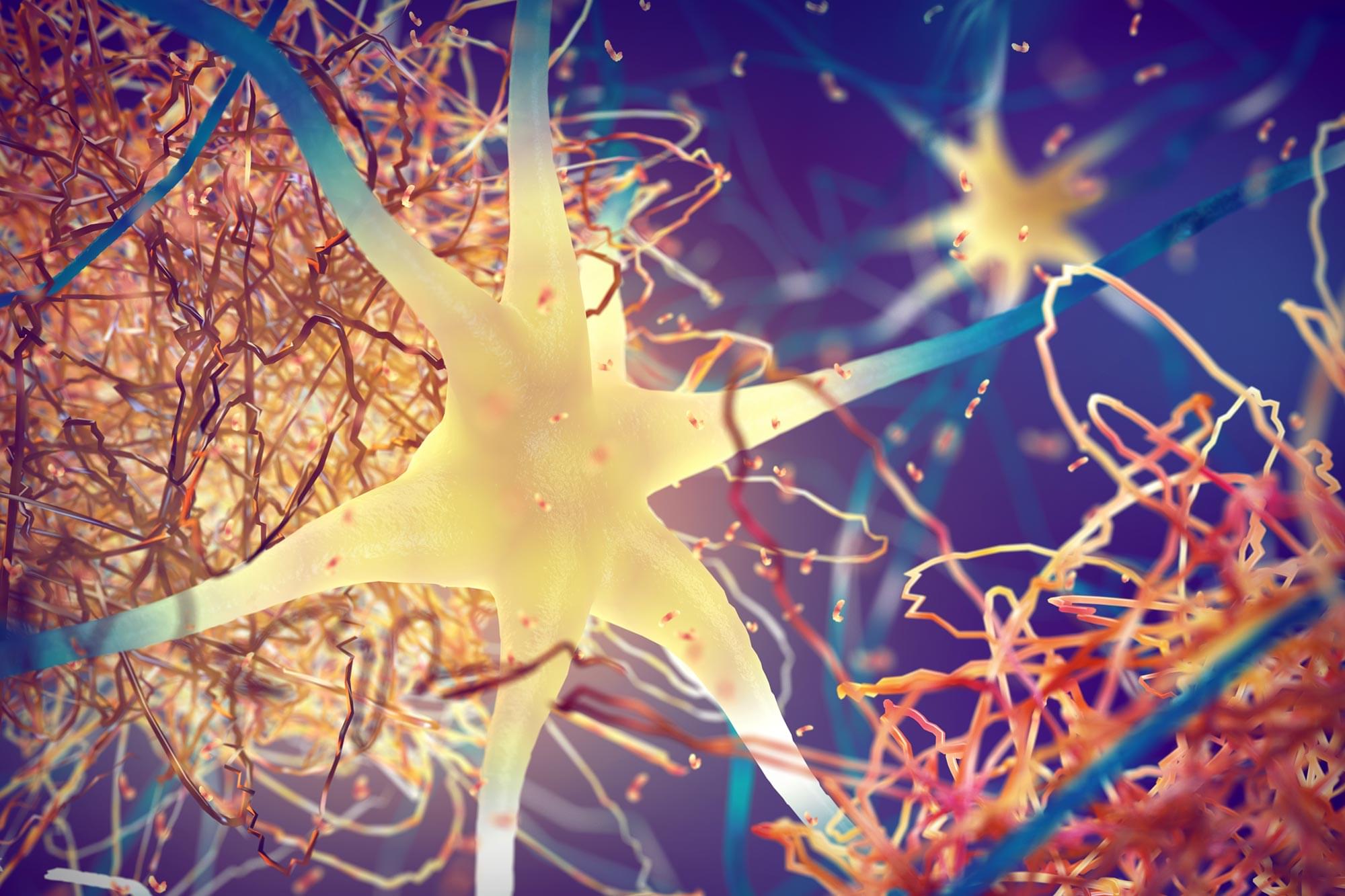
This study describes a novel ATXN2 expansion within the classic pathogenic range for spinocerebellar ataxia 2 that manifests as an early-onset neurodegenerative disorder in the homozygous state, while being asymptomatic into late adulthood in the heterozygous state.


The length and content of ATXN2 trinucleotide repeat significantly influences disease development and clinical phenotype. ATXN2 alleles containing 13–31 CAG trinucleotide repeats are normal and commonly found in healthy individuals4 and over 90% of tested individuals possess an allele containing 22 CAG repeats.21 Spinocerebellar ataxia type 2 is caused by dominant alleles of 33 or more CAG trinucleotide repeats.11,22 Alleles containing 33–34 CAG repeats are considered reduced penetrance alleles, and carriers may or may not develop late onset ataxia.22 Fully penetrant alleles most commonly have 37–39 CAG repeats and are pathogenic for SCA2.11 While SCA2 alleles of 31 pure CAG repeats exhibit high instability on inheritance, it has been proposed that CAA interruptions confers meiotic stability.23 An anticipation phenomenon in SCA2 has also been described, consisting of earlier disease onset and increased clinical severity in subsequent generations which are mirrored by an increase in CAG repeat size.12 Patients with SCA2-related parkinsonism carry intermediate range alleles and possess alleles with CAA interruptions.24,25 Similarly, ATNX2 variants associated with ALS are CAA interrupted and are rarely in the pathogenic range of SCA2.26,27 Contrasting with trinucleotide expansion diseases, repeat size has no bearing on ALS AO but correlates with disease risk.28 ATXN2 has been identified as a disease modifier gene for a variety of neurologic conditions and similarly, various genes may influence the AO of SCA2, including long normal repeats in the CACNA1A and RAI1 genes.29 Nonetheless, the most important predictor of AO and clinical severity remains the polyglutamine repeat expansion size.30

Infantile and childhood forms of SCA2 are described, and these patients present with a multi-systematic neurodegenerative disorder including developmental delay, retinitis pigmentosa, optic atrophy, hypotonia, seizures, facial dysmorphism, dystonic features, and early mortality.21,31 Infantile cases all possess extreme length CAG repeats (range 69–884) in the heterozygous state, with clinical severity related to repeat size, and inherited with an anticipation phenomenon from parents within the fully penetrant range of SCA2 (range 39–47 CAG repeats).21,31

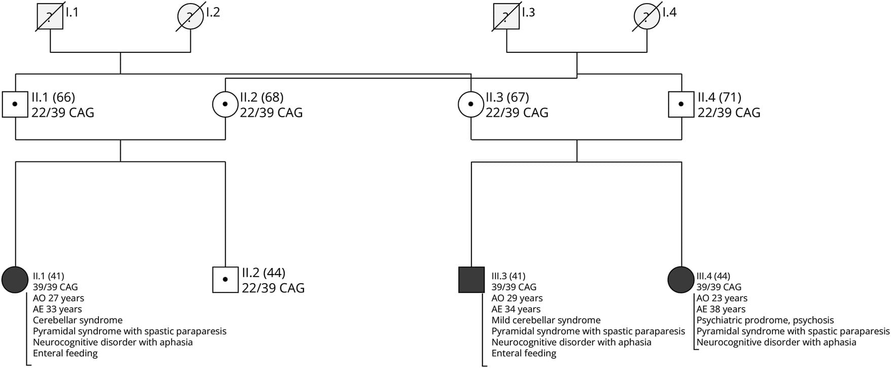
Homozygous cases of SCA2 are exceedingly rare.32,33 Notably, a patient with 31/31 CAG alleles developed late-onset cerebellar ataxia, suggesting that patients with homozygous variants may manifest signs of disease within a nonpathogenic variant range, that is not associated with disease development in the heterozygous state.18,32 Two homozygous cases from an Indian family with 35/37 and 36/39 CAG repeats alleles developed early onset, levodopa responsive Parkinson disease without ataxia,33 while several family members with heterozygous ATXN2 variants exhibited parkinsonism and/or ataxia with variable ages of onset ranging from adulthood to their sixties.33 Moreover, two homozygous cases with intermediate alleles of 32/3217 and 33/3327 displayed a pure ALS phenotype, without ataxia. These cases highlight the phenotypical variability of homozygous ATXN2 variants.

Affecting a Signaling Pathway Alleviates Alzheimer’s in Mice

A new study shows that the overexpression of somatostatin (SST), a neuropeptide produced in neurons and acting mostly on microglia, lowers inflammation and amyloid β burden, improving cognitive abilities in a mouse model of Alzheimer’s. Drugs affecting this pathway are already available [1].

The unusual suspect

In Alzheimer’s disease, many signaling pathways in the brain become dysregulated. Since going after the main hallmarks of the disease (amyloid β and tau protein accumulation) has only yielded modest results so far, scientists are exploring various secondary targets whose levels correlate with the disease.

Single-cell epigenomics uncovers heterochromatin instability and transcription factor dysfunction during mouse brain aging

Amaral et al. present a single-cell atlas of brain aging, revealing coordinated chromatin and gene expression changes across multiple regions from young to old mice. Their analyses show that aging involves loss of progenitor cells, dysregulation of master transcription factors, and destabilization of heterochromatin, driving a gradual erosion of cellular identity.

/* */