Toggle light / dark theme

Link :


Glioblastoma, an aggressive and often fatal form of brain cancer, has long posed a formidable challenge to doctors and patients alike. Yet, a groundbreaking clinical trial is offering a glimmer of hope, capturing global attention for its potential to revolutionize cancer treatment. A 62-year-old engineer, faced with a grim prognosis, has experienced something extraordinary—his tumour has shrunk significantly in a matter of weeks. This remarkable outcome marks the beginning of a journey that could redefine how we treat one of the most challenging cancers. What makes this approach so promising, and how could it change the future for patients?

Glioblastoma, often referred to as glioblastoma multiforme (GBM), is the most aggressive and common form of primary brain cancer in adults. Originating from glial cells—specifically astrocytes that support nerve cells—this malignancy is notorious for its rapid growth and diffuse infiltration into surrounding brain tissue, making complete surgical removal challenging.

While research continues on the potential of psychedelics as a clinical treatment, a recent study highlights the need to better understand their adverse effects.


Subscribe Now / Learn More

PsychiatryOnline subscription options offer access to the DSM-5-TR® library, books, journals, CME, and patient resources. This all-in-one virtual library provides psychiatrists and mental health professionals with key resources for diagnosis, treatment, research, and professional development.

Need more help? PsychiatryOnline Customer Service may be reached by emailing [email protected] or by calling 800−368−5777 (in the U.S.) or 703−907−7322 (outside the U.S.).

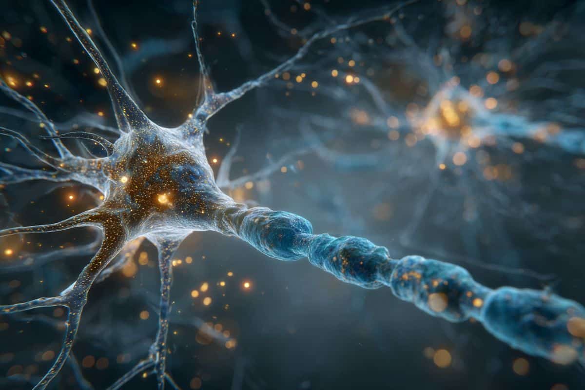
Many cells in our body have a single primary cilium, a micrometer-long, hair-like organelle protruding from the that transmits cellular signals. Cilia are important for regulating cellular processes, but because of their small size and number, it has been difficult for scientists to explore cilia in brain cells with traditional techniques, leaving their organization and function unclear.

In a series of papers appearing in Current Biology, the Journal of Cell Biology, and the Proceedings of the National Academy of Sciences, researchers at HHMI’s Janelia Research Campus, the Allen Institute, the University of Texas Southwestern Medical Center, and Harvard Medical School used super high-resolution 3D electron microscopy images of mouse brain tissue generated for creating connectomes to get the best look yet at .

Researchers have developed a reliable and reproducible way to fabricate tapered polymer optical fibers that can be used to deliver light to the brain. These fibers could be used in animal studies to help scientists better understand treatments and interventions for various neurological conditions.

The tapered fibers are optimized for neuroscience research techniques, such as optogenetic experiments and fiber photometry, which rely on the interaction between genetically modified neurons and delivered to and/or collected from the .

“Unlike standard optical fibers, which are cylindrical, the tapered fibers we developed have a conical shape, which allows them to penetrate the tissue with more ease and to deliver light to larger volumes of the brain,” said research team member Marcello Meneghetti from the Neural Devices and Gas Photonics group at the Technical University of Denmark.

Researchers discovered that electrical synapses filter sensory signals in animals, enabling context-specific decision-making—a finding with broad implications for neuroscience.

Scientists from Yale University

Established in 1701, Yale University is a private Ivy League research university in New Haven, Connecticut. It is the third-oldest institution of higher education in the United States and is organized into fourteen constituent schools: the original undergraduate college, the Yale Graduate School of Arts and Sciences and twelve professional schools. It is named after British East India Company governor Elihu Yale.

The coordinated activity of brain cells, like birds flying in formation, helps us behave intelligently in new situations, according to a study led by Cedars-Sinai investigators. The work, published in the peer-reviewed journal Nature, is the first to illuminate the neurological processes known as abstraction and inference in the human brain.

“Abstraction allows us to ignore irrelevant details and focus on the information we need in order to act, and inference is the use of knowledge to make educated guesses about the world around us,” said Ueli Rutishauser, PhD, professor and Board of Governors Chair in Neurosciences at Cedars-Sinai and co-corresponding author of the study. “Both are important parts of cognition and learning.”

Humans often use these two cognitive processes together to rapidly learn about and act appropriately in new environments. One example of this is an American driver who rents a car in London for the first time.

Gamma oscillations in the brain reveal pain intensity, driven by PV interneurons in the somatosensory cortex. New research highlights their role as biomarkers and therapeutic targets for pain management.


Summary: Parvalbumin (PV) interneurons in the primary somatosensory cortex (S1) have been identified as key players in encoding pain intensity and driving gamma oscillations, according to a study. Cross-species experiments confirmed that gamma oscillations in S1 selectively reflect pain levels in humans and are linked to PV interneuron activity in rodents.

Optogenetic manipulation of these interneurons demonstrated their ability to modulate pain-related behaviors, solidifying their role in pain processing. The findings establish a direct connection between PV interneurons and gamma oscillations, highlighting their potential as a biomarker and target for pain therapies.

Researchers have discovered that neurotransmitters like dopamine.

Dopamine is a crucial neurotransmitter involved in many important functions in the brain, particularly those related to pleasure, reward, motivation, and motor control. It plays a central role in the brain’s reward system, where it helps reinforce rewarding behaviors by increasing pleasure and satisfaction, making it critical for habit formation and addictive behaviors. Dopamine is also vital for regulating movement, and deficiencies in dopamine production are linked to neurological disorders such as Parkinson’s disease. Additionally, dopamine influences various other functions, including mood regulation, learning, and attention, making it a key focus in studies of both mental health and neurodegenerative diseases.

New research uncovers how neuropilin2 gene mutations disrupt brain balance, linking inhibitory neuron migration to autism and epilepsy. Study offers insights for targeted therapies.


Source: UCR

The gene neuropilin2 encodes a receptor involved in cell-cell interactions in the brain and plays a key role in regulating the development of neural circuits.

Neuropilin2 controls migration of inhibitory neurons as well as the formation and maintenance of synaptic connections in excitatory neurons — two crucial components of brain activity.