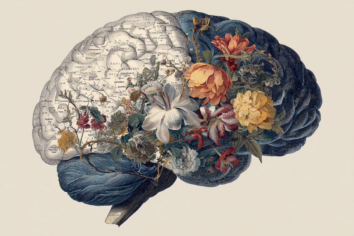
Researchers identify the rostral prefrontal cortex as the essential bridge between spontaneous and executive brain networks that drives creativity.
Become a Big Think member to unlock expert classes, premium print issues, exclusive events and more: https://bigthink.com/membership/?utm_… How your biology and environment make your decisions for you, according to Dr. Robert Sapolsky.
Up next, Your reptilian brain, explained ► • Your reptilian brain, explained | Robert S…
Robert Sapolsky, PhD is an author, researcher, and professor of biology, neurology, and neurosurgery at Stanford University. In this interview with Big Think’s Editor-in-Chief, Robert Chapman Smith, Sapolsky discusses the content of his most recent book, “Determined: The Science of Life Without Free Will.”
Being held as a child, growing up in a collectivist culture, or experiencing any sort of brain trauma – among hundreds of other things – can shape your internal biases and ultimately influence the decisions you make. This, explains Sapolsky, means that free will is not – and never has been – real. Even physiological factors like hunger can discreetly influence decision making, as discovered in a study that found judges were more likely to grant parole after they had eaten.
This insight is key for interpreting human behavior, helping not only scientists but those who aim to evolve education systems, mental health research, and even policy making.
Go Deeper with Big Think:
Is there a quantum reason we could have free will? Neil deGrasse Tyson and comedian Chuck Nice explore the concept of free will and predetermination with neuroscientist, biologist, and author of Determined: The Science of Life Without Free Will, Robert Sapolsky.
A special thanks from our editors to Robert Sapolsky’s dog.
Could we put an end to the question of whether or not we have free will? Discover “The Hungry Judge Effect” and how little bits of biology affect our actions. We break down a physicist’s perspective of free will, The Big Bang, and chaos theory. Is it enough to just feel like we have free will? Why is it an issue to think you have free will if you don’t?
We discuss the difference between free will in big decisions versus everyday decisions. How do you turn out to be the type of person who chooses vanilla ice cream over strawberry? We explore how quantum physics and virtual particles factor into predetermination. Could quantum randomness change the actions of an atom? How can society best account for a lack of free will? Are people still responsible for their actions?
What would Chuck do if he could do anything he wanted? We also discuss the benefits of a society that acknowledges powers outside of our control and scientific advancements made. How is meritocracy impacted by free will? Plus, can you change if people believe in free will if they have no free will in believing so?
Thanks to our Patrons Pro Handyman, Brad K. Daniels, Starman, Stephen Somers, Nina Kane, Paul Applegate, and David Goldberg for supporting us this week.
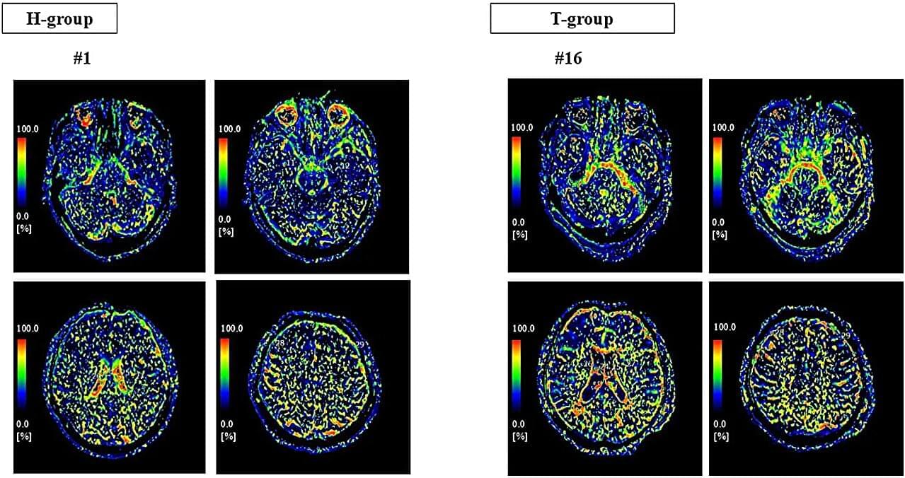
Researchers at University of Tsukuba have found that cerebrospinal fluid (CSF) microdynamic motion shows region-specific alterations after mild traumatic brain injury (TBI). Using a specialized magnetic resonance imaging (MRI) technique, the team noninvasively visualized these CSF changes, which have been difficult to quantify with conventional imaging. The approach is expected to advance the understanding of the relationship between post-traumatic brain conditions and cognitive function. The study is published in Frontiers in Neuroscience.
The brain contains cerebrospinal fluid (CSF), which protects neural tissue and helps clear metabolic waste. Rather than being static, CSF exhibits continuous subtle motion, and this motion is thought to be closely linked to brain health. However, little has been known about how CSF motion is altered after a mild head injury.
The researchers employed a specialized magnetic resonance imaging (MRI) technique known as intravoxel incoherent motion (IVIM) MRI to evaluate CSF microdynamic motion through the incoherent movement of water molecules. The results showed that, after mild traumatic brain injury (TBI), CSF motion increased in some brain regions and decreased in others.
Epilepsy is best known for seizures, but many people with the condition also experience much more frequent and subtler disruptions. These brief bursts of abnormal brain activity, called interictal epileptiform discharges (IEDs), can happen thousands of times a day, interfering with attention, memory, language, and sleep.
Scientists at UC San Francisco have discovered that these “brain blips” are not random events, as had been believed. Rather, they unfold in a predictable pattern that can be detected a full second before they occur — raising new possibilities to ward them off altogether.
The researchers used a high-resolution technology recently adapted for humans that can record the activity of individual neurons. They tracked more than 1,000 neurons in four patients undergoing surgery for epilepsy.
Most physicists are materialists who believe the world consists of physical particles at the fundamental level. Others have argued reality is a simulation or a hallucination of the brain. But Andrew T. Jaffe challenges all of these views, proposing an alternative consciousness-first theory where space and time arise as within a dream.
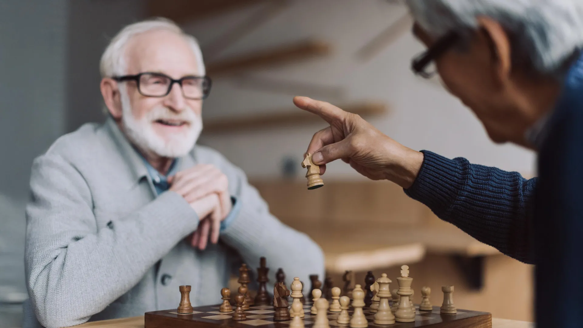
These individuals consistently perform on memory tests at levels similar to people at least 30 years younger, challenging the long-standing belief that cognitive decline is unavoidable with age.
Over decades of research, scientists have noticed some lifestyle and personality traits that set SuperAgers apart from their peers, including being highly social and outgoing. Still, the most surprising discoveries have come from examining their brains. “It’s really what we’ve found in their brains that’s been so earth-shattering for us,” said Dr. Sandra Weintraub, a professor of psychiatry and behavioral sciences and neurology at Northwestern University Feinberg School of Medicine.
By identifying both biological and behavioral patterns linked to SuperAging, researchers hope to develop new approaches to strengthen cognitive resilience and reduce the risk of Alzheimer’s disease and other forms of dementia.
Our speaker this month is Jordan Sparks with the Sparks Brain Preservation organization in Oregon. Our event is in ZOOM Only, no in person meeting this month, meeting ins ZOOM on Thursday, April 30th, opening at 6:00 PM for our social hour, with the main event starting at 7:00 PM Eastern Time Jordan will tell us about his project, which was formerly the Oregon Brain Preservation, and before that Jordan formed Oregon Cryonics. This is an entirely different type of bio-stasis then cryonics. Their stated goal is to preserve the structure of the entire brain at a fine ultrastructural level. This includes the synaptic architecture as well as detailed molecular information such as protein post-translational modifications, cellular epigenetic patterns, and subcellular distributions of molecules.
By mapping all the possible variations in a single gene, researchers have uncovered a previously hidden neurodevelopmental condition.
ReNU syndrome is a rare, inherited neurodevelopmental disorder identified in 2024 that affects brain function, development, and motor skills and is predicted to affect tens of thousands of individuals worldwide.