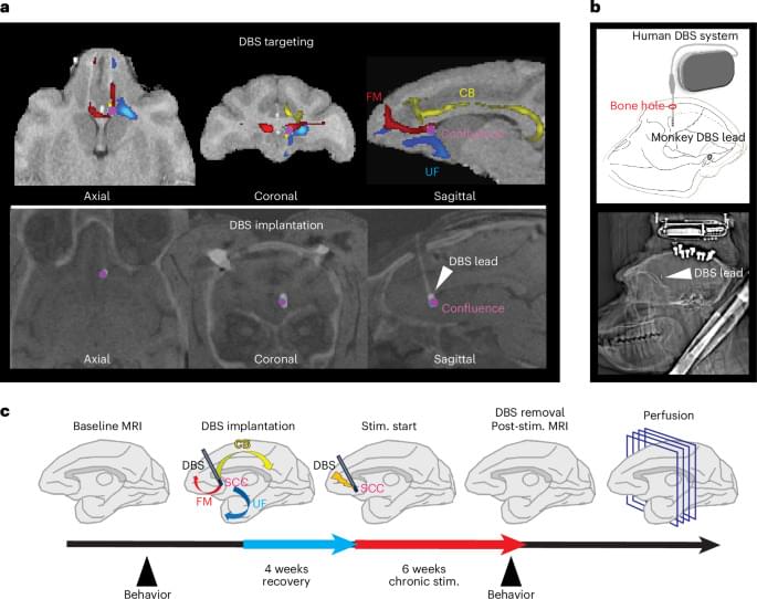
In a nonhuman primate model, Fujimoto et al. show that deep brain stimulation promotes white matter remodeling and reorganizes brain-wide functional networks, detailing a mechanism through which this neuromodulation therapy may treat depression.

Smaller plastic particles have more effects on neurons, the key information processing cells of the brain, new research from the University of Eastern Finland shows. In the study, neuronal cells were exposed to polystyrene nanoplastics at low doses to study subtle changes.
Plastic production continues to rise, despite worldwide concerns. In addition to environmental implications, there is an increasing interest in how exposure to plastics may impact human health, but our understanding is still limited. Only recently it was shown that plastics can accumulate also in the human brain.
Plastic particles smaller than 5,000 nm in diameter are called microplastics, and the smallest plastic particles with a diameter of less than 1,000 nm are called nanoplastics. The small size of nanoplastics enables them to interact with various cell types, and other particles or biological mass, such as bacteria. Compared to microplastics, nanoplastics have larger adsorption capacity and penetrate through biological barriers more easily. This makes them potentially more harmful and a compelling target for research in the field of neurobiology.
ATT Business: Switch to AT&T Business at business.att.com.
Shopify: Sign up for your one-dollar-per-month trial period at https://shopify.com/impact.
Welcome back to Impact Theory with Tom Bilyeu. In today’s episode, Tom Bilyeu dives deep into one of the most provocative questions facing science and philosophy: Do we really have free will, or are we all just highly sophisticated NPCs—non-player characters—running a program inside a vast, resource-efficient simulation? Drawing on groundbreaking neuroscience experiments, the story of Phineas Gage, quantum mechanics, and the work of leading thinkers like Robert Sapolsky, Tom Bilyeu challenges everything we think we know about choice, consciousness, and the true nature of reality.
But this isn’t an episode about nihilism. Instead, Tom Bilyeu reveals why embracing the truth of a stochastically deterministic universe can actually make life feel more meaningful, freeing us from the weight of the past and inspiring us to make the most of every moment—programmed or not. Get ready to question your assumptions and see the world from a whole new perspective.
00:00 — Intro.
01:38 — Part 1: It’s Biology All The Way Down.
14:22 — Part 2: Quantum Mechanics Bury the Notion of Free Will.
19:53 — Part 3: The Last Hiding Places of Free Will.
27:51 — Part 4: Why Being An NPC Is The Best News You’ll Ever Get.
Sign up for my AI Masterclass: https://tombilyeu.com/ai-masterclass.
Check us out wherever you get your podcasts:
Spotify: https://open.spotify.com/show/1nARKz2…
Apple: https://podcasts.apple.com/us/podcast…
Do you need my help?
STARTING a business: join me here inside ZERO TO FOUNDER (https://tombilyeu.com/zero-to-founder)
Get the exact systems, mindset shifts, and principles that built a $1B brand delivered straight to your inbox every week. Subscribe for free (https://tombilyeu.com)
Check out our Video game — Project Kyzen: (https://projectkyzen.io/)
Catch Me Streaming on Twitch — (/ tombilyeu)
Link to IT discord: / discord.
Tom’s Favorite Things List: https://amzn.to/41Ftt7e.
FOLLOW TOM:
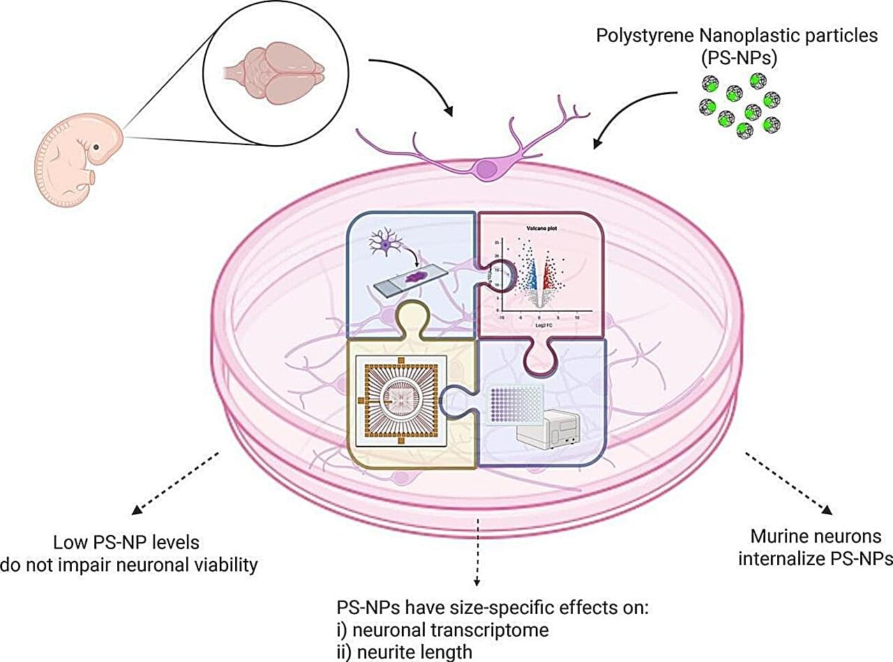
Smaller plastic particles have more effects on neurons, the key information processing cells of the brain, new research from the University of Eastern Finland shows. In the study, neuronal cells were exposed to polystyrene nanoplastics at low doses to study subtle changes.
The study is published in the journal NanoImpact.
Plastic production continues to rise, despite worldwide concerns. In addition to environmental implications, there is an increasing interest in how exposure to plastics may impact human health, but our understanding is still limited. Only recently was it shown that plastics can also accumulate in the human brain.
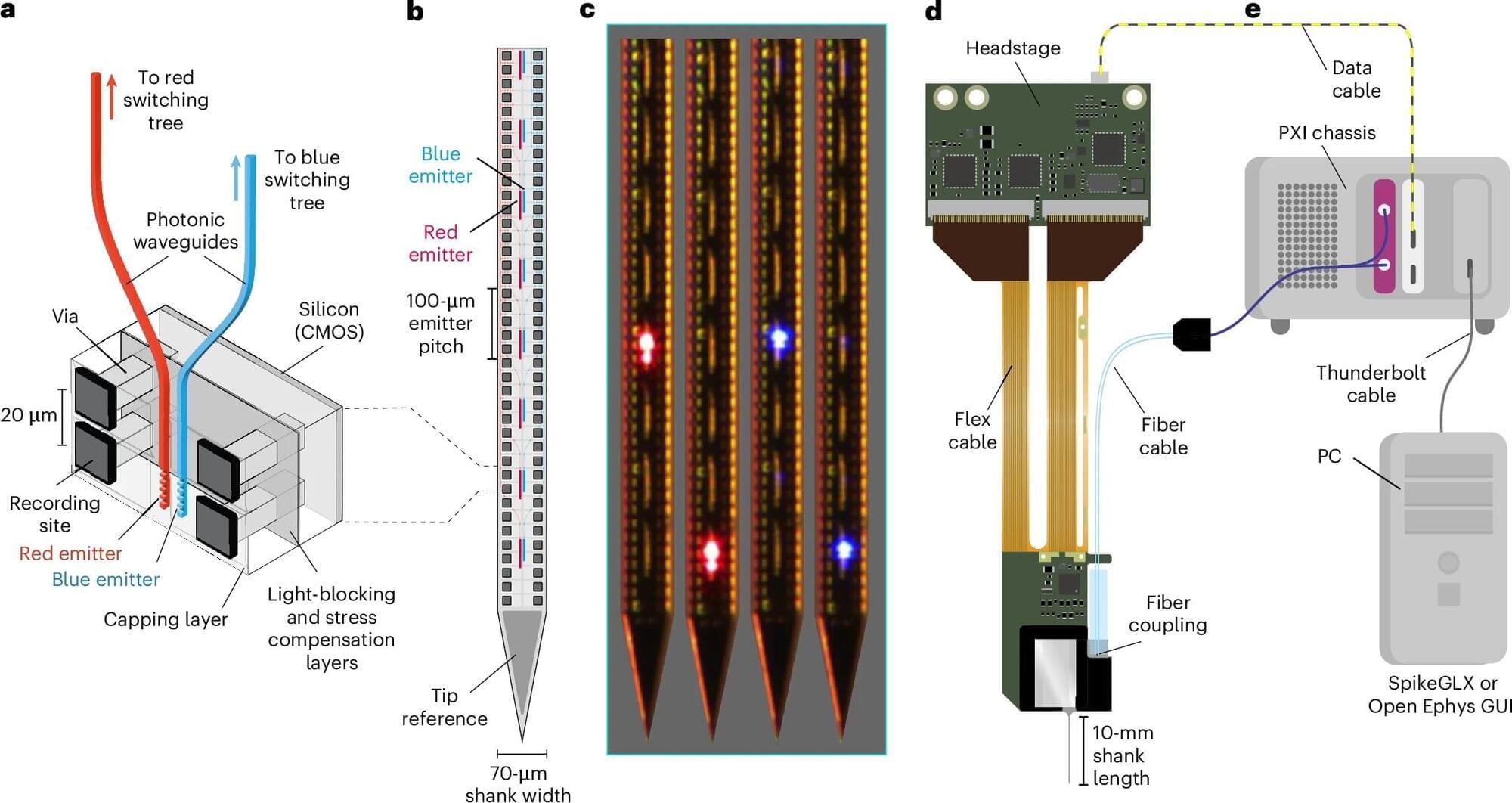
A new breakthrough technology, co-developed by UCL scientists, that simultaneously records and manipulates neuron activity deep within the brain could transform our understanding of neural circuits and neurological conditions, such as Alzheimer’s disease and schizophrenia.
The device, known as Neuropixels Opto and researched in mice, integrates two powerful but traditionally separate techniques—electrophysiology (the study of the electrical activity of living cells) and optogenetics (combining genetics and optics to control cells). They form a single probe, enabling unprecedented insight into how individual neurons in the brain function and interact.
Published in Nature Methods, the system allows researchers to monitor the electrical activity of hundreds of neurons while also selectively activating or silencing specific cells using light.
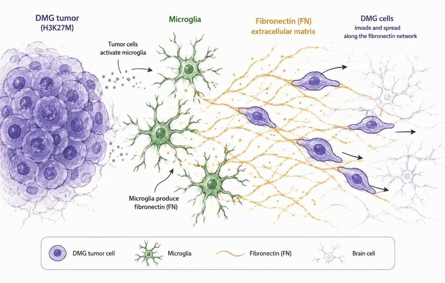
Researchers at the Institute of Environmental Medicine (IMM), Karolinska Institutet, have identified a possible mechanism behind the spread of the aggressive brain tumor diffuse midline glioma. The study shows that the brain’s own immune cells, microglia, may contribute to the tumor’s invasive capacity by producing the protein fibronectin. The results are published in the journal Cell Death & Disease.
Diffuse midline glioma (DMG), also known as diffuse intrinsic pontine glioma (DIPG), is a rare but highly aggressive brain tumor that primarily affects children. There is currently no effective treatment, and the prognosis is very poor.
In the present study, the researchers investigated how microglia—the brain’s immune cells—are affected by tumor cells and what role they play in disease progression.