To learn more, please visit the YouTube Help Center: https://www.youtube.com/help
Category: neuroscience – Page 20
Frontiers: For nearly a decade, the idea that ‘the body keeps the score’ has shaped public and clinical understanding of trauma (van der Kolk, 2014)
It is an enticing metaphor—implying that experience is literally inscribed in flesh, that the body bears the scars of what the mind cannot face. Yet recent advances in computational and systems neuroscience reveal that this image, while emotionally compelling, is biologically inaccurate. The body proper does not store trauma; the brain dynamically reenacts it through maladaptive inference. What endures after trauma is not a memory lodged in non-innervated tissue but a collapse of flexibility—a loss of metastability, the brain’s ability to fluidly switch among semi-stable network states.
In computational terms, trauma over-weights the precision of danger priors: the brain assigns excessive confidence to threat predictions, constraining inference based on the prior premise of enduring danger. The result is hypervigilance, flashbacks, and avoidance—symptoms of a system caught in self-confirming predictions. Mathematically, this overconfidence means one cannot escape local minima—in a free energy landscape—that become deeply and precisely engrained (i.e., trapped in a ravine with steep sides, where precision corresponds to the local curvature or steepness).
This rigidity contrasts with a healthy brain’s metastable dynamics, where neuronal networks continually integrate and segregate in response to context. This allows neuronal dynamics to explore multiple (unstable) interpretations of the world. Hellyer and colleagues demonstrated that metastability is a hallmark of cognitive flexibility: the capacity for neural coalitions to assemble transiently and adapt quickly. Using both empirical and computational approaches, Hellyer et al. (2015) showed that reduced metastability arising from damage to the structural connectome was associated with diminished cognitive flexibility and impaired information processing. Trauma erodes this fluidity, trapping the brain in narrow basins of fear and defensive salience. To restore mental health is not about ‘releasing’ stored emotion but reestablishing dynamic equilibrium enabling the brain’s ability to move with graceful agility over a landscape of beliefs, commitments and intentions.
Third electrode pair can sharpen deep brain stimulation technique, mouse experiments suggest
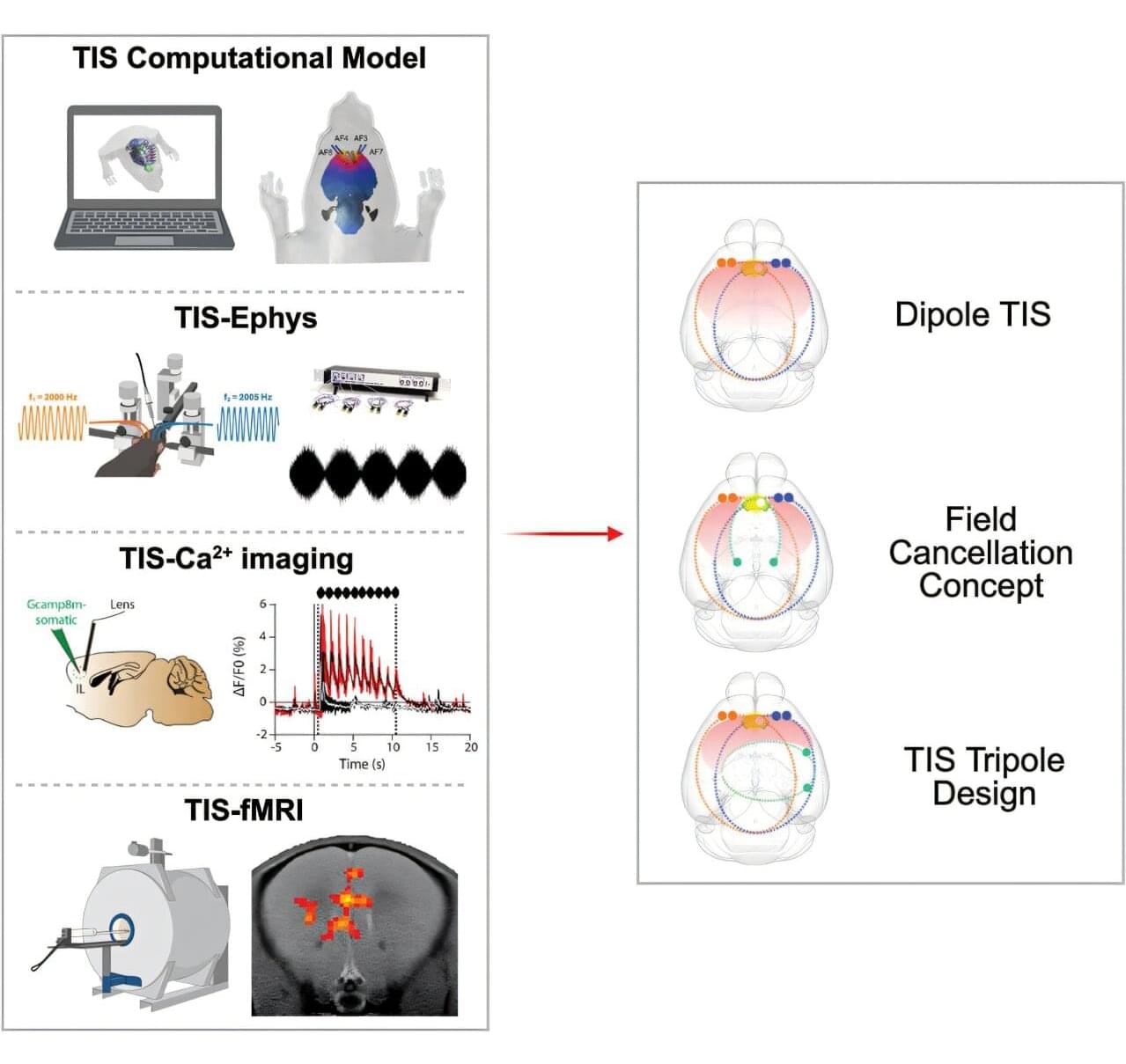
A study by UNIGE, in collaboration with ETH Zurich, has significantly improved the accuracy of a noninvasive brain stimulation technique, paving the way for its use in the treatment of neurological and psychiatric disorders.
Brain stimulation techniques can correct abnormal activity in the neural circuits involved in conditions such as Parkinson’s disease and depression. However, current transcranial stimulation methods delivered through the scalp reach only the brain’s surface, limiting their effectiveness. Deep brain stimulation, on the other hand, can target deeper structures but requires surgical implantation of electrodes.
A team from the Synapsy Center for Neuroscience and Mental Health Research at the University of Geneva (UNIGE), in collaboration with ETH Zurich, the Wyss Center Geneva and EPFL, has improved a promising intermediate technology called “temporal interference stimulation.” This method could allow deeper and more targeted noninvasive brain stimulation. The study is published in Cell Systems.
China Just Beat Elon Musk With A Chip Inside The Human Brain
🔒 Stay private online with NordVPN →
https://go.nordvpn.net/SHBMd.
Beat censorship and tracking. 30-day money-back guarantee.
Affiliate link — I earn a small commission at no cost to you.
A major new development in brain-computer technology is raising eyebrows across the tech world. While Elon Musk’s Neuralink has dominated headlines for years, a breakthrough emerging from China is now sparking fresh debate about who is really leading the race to connect the human brain with advanced computing systems.
In this video, we take a closer look at the latest brain-chip innovation, what makes it different from existing neural interface projects, and why experts are paying close attention. As competition intensifies between global technology powers, advances in neural implants could reshape medicine, communication, and even the future relationship between humans and machines.
Could this new achievement challenge Neuralink’s position at the center of the brain-tech conversation? And what does it mean for the future of artificial intelligence, neuroscience, and human enhancement? The implications may be far bigger than many people realize.
Memory reactivation underlies experience-dependent adaptive regulation of sleep
When we fall asleep, our brains don’t just shut off; they get to work. One of their primary jobs is memory consolidation—sorting through the events of the day and filing them into long-term storage. The brain does this by spontaneously “reactivating” or replaying memories.
Recent memories are consolidated during sleep by spontaneous reactivation. However, whether and how memory reactivation affects sleep dynamics remain unclear. By tracking and modulating memory activity during sleep in mice, we revealed that negative memory reactivation promoted arousal, whereas positive memory supported sleep stability. This regulation was mediated by the reactivation of experience-specific hippocampus-amygdala engram circuits during sleep. In chronic stress models, negative memory reactivation promoted sleep disturbance, and targeted suppression of memory reactivation restored normal sleep. Our findings establish a memory-dependent sleep regulation in which memory reactivation engages downstream circuits responsive to specific memory content.
Organoid Intelligence | Dr. Thomas Hartung | XPANSE 2024
Delve into the fascinating world of organoid intelligence at XPANSE 2024 in Abu Dhabi. Presented by Dr. Thomas Hartung, Professor of Medical Microbiology at Johns Hopkins University, this session explores the cutting-edge research and potential of lab-grown organoids to revolutionize computing, medicine, and neuroscience.
XPANSE, the world’s first visioning of the future with exponential technologies, is an Abu Dhabi-based global initiative and an invitation-only forum for exponential technology. XPANSE 2024, hosted by ADQ, convened 3,000 world’s brightest minds, technology trailblazers, Nobel Laureates, industry leaders, CEOs, ministers and scientists to set the horizons of exponential technologies spanning quantum, genomics, exotic computing, embodied intelligence, next-gen 2D matter, AGI, Brain-Machine Interfaces, Future G and beyond.
Be the first one to know about XPANSE 2025 ►► https://mailchi.mp/xpanse.world/sign–… connected with our community & get insider insights ►► / xpanse-world Follow XPANSE on Instagram ►►
/ xpanseworld Follow XPANSE on X ►► https://twitter.com/XPANSEWORLD
Stay connected with our community & get insider insights ►► / xpanse-world.
Follow XPANSE on Instagram ►► / xpanseworld.
Follow XPANSE on X ►►https://twitter.com/XPANSEWORLD
New Discoveries Challenge Everything We Knew About Brain Evolution
Support this channel on Patreon to help me make this a full time job: / whatdamath (Unreleased videos, extra footage, DMs, no ads)
Alternatively, PayPal donations can be sent here: http://paypal.me/whatdamath.
Get a Wonderful Person Tee: https://teespring.com/stores/whatdamath.
More cool designs are on Amazon: https://amzn.to/3QFIrFX
Hello and welcome! My name is Anton and in this video, we will talk about a few studies that explain how the human brain developed complexity.
Links:
https://linkinghub.elsevier.com/retri…
Other videos:
• Surprise Evidence That Gut Microbes Direct…
• Mindblowing Discoveries About Bacteria Liv…
• Direct Connection Between Gut Microbiome a…
#brain #biology #evolution.
0:00 Discoveries about the evolution of the brain.
1:20 800 Million years ago… how it all began.
3:10 Did nervous system evolve multiple times? Comb jellies.
4:45 Big brains — primates vs octopuses.
9:20 Human brains and human intelligence genes.
11:20 Gut microbes and fuel for the brain.
12:20 Conclusions and implications.
Enjoy and please subscribe.
Bitcoin/Ethereum to spare? Donate them here to help this channel grow!
bc1qnkl3nk0zt7w0xzrgur9pnkcduj7a3xxllcn7d4
or ETH: 0x60f088B10b03115405d313f964BeA93eF0Bd3DbF
The hardware used to record these videos:
Redefining Matter | Why Matter Is Not What We Think It Is?
What is matter, really? Is matter an independent substance, or is reality fundamentally relational? In this episode, we explore some of the deepest questions in philosophy, metaphysics, and modern science, including Quantum Physics, Relativity, Quantum Field Theory, Dark Matter, Consciousness, Space, Time, Cosmology, and the Nature of Reality itself.
From atoms and particles to galaxies and the Universe, modern science increasingly points toward a world of processes, relationships, and dynamic structures rather than isolated objects. Could Matter and Consciousness be different expressions of the same underlying Reality? What can Systems Thinking, Complexity Theory, Nonduality, Taoism, Buddhism, and Vedanta contribute to our understanding of existence?
Let us examine the Nature of Matter, the mystery of Dark Matter, the meaning of Space-Time, and the interconnected fabric of the cosmos. This exploration may challenge the way you think about Reality, Existence, Consciousness, and your place within the Universe.
#QuantumPhysics #Consciousness #NatureOfReality #WhatIsMatter #Relativity #QuantumFieldTheory #DarkMatter #Universe #Cosmology #Philosophy #Metaphysics #ScienceAndPhilosophy #NonDuality #Taoism #Buddhism #Vedanta #SystemsThinking #ComplexityTheory #Interconnectedness #meaningoflife.
0:00 Intro.
0:55 A Necessary Correction of Attitude.
4:39 What is Matter?
8:09 Rethinking Properties.
10:34 An Important Question.
14:11 Redefining Matter.
17:43 Outro.
If you love my content, you can support me here: https://buymeacoffee.com/philosophydi… For inquiries: [email protected] ============================= 🎬Suggested videos for you: ▶️ • 3 Quantum Entanglement ▶️
• 2 Wave-Particle Duality ▶️
• 1 Observer Effect ▶️
• Food and Your Mind | How What You Eat Shap… ▶️
• Indian Vegetarian Cooking | How to Make De… ▶️
• Merry Christmas 🎄 ▶️
• 2 What does addiction feel like? ▶️
• Quickly cooking Chinese food😋 ▶️
• 1 Do we really need the “shortcut” to spi… ▶️
• 4 Re-understanding Manifestation ▶️
• 3 Re-understanding Matter ▶️
• 2 Re-understanding Energy ▶️
• 7 Where Does Existence Come From? Final An… ▶️
• 6 There was no “Creation” =================================.
The Past, Present, & Future of Brain-Computer Interfaces By Rolando Masís-Obando
In brief: A historical look into how brain computer interfaces have transformed over the past few decades: the landmark research of the past, the landmark research of today, and how it’s going to transform the future of XR. As a neuroscientist for about a decade, my work has focused on how people represent spatial contexts, concepts, and events. I have been able to place people in VR experiences and then through the use of neuroimaging and AI methods untangle their thoughts and how those thoughts influenced what they remember. As this neuroimaging technology reduces in form-factor and increases in accessibility, we can no longer turn a blind-eye to how it may be used nefariously in consumer products. In this talk, I will describe how Brain-computer interfaces (BCIs) have been defined over the years, how research in this field has catapulted, an overview of the neuroscience behind the technology, the landmark studies of the past and present, use-cases in which XR, robotics, prosthetics and BCI have intertwined, and how new AI models are being used to perform mind-reading of both language and mental images. “With great power comes great responsibility” – I will end the talk by describing how and what it means for the future of XR and why it’s important to be careful with this technology, but also how incredibly empowering it can be for the future of XR.
Scientists Put a Fruit Fly’s Brain in a Computer Simulation… What It Did Is Now Scaring Scientists
Scientists have achieved an incredible breakthrough by recreating the brain of a fruit fly inside a computer simulation. By mapping around 140,000 neurons and millions of connections, they built a digital brain that can sense its environment, process information, and even control a virtual body. In the simulation, the digital fly was able to search for food, respond to stimuli, and show behaviors that were not directly programmed by scientists. This discovery shows how powerful neural connections are in generating behavior. It also raises fascinating questions about the nature of intelligence, consciousness, and whether complex brains—including ours—could one day be simulated in computers.
sources
https://eon.systems/updates/embodied-brain-emulation.
Research Paper for more information.
https://marginalrevolution.com/margin…
#Science.
#Neuroscience.
#ArtificialIntelligence.
#BrainSimulation.
#FruitFlyBrain.
#Connectome.
#FutureTech.
#ComputerSimulation.
#NeuralNetworks.
#ScienceDiscovery